Diagnosis of Scalp Rashes: Clinical Approach
Master scalp rash diagnosis with clinical examination, history, and diagnostic testing.

Diagnosis of Scalp Rashes: A Clinical Approach
Scalp rashes represent a diverse range of dermatological conditions that require systematic evaluation and careful clinical assessment. When patients present with complaints of scalp problems, healthcare providers must conduct a thorough evaluation that extends beyond the scalp itself. A comprehensive approach involving detailed medical history, focused cutaneous examination, and appropriate diagnostic testing is essential for accurate diagnosis and effective management.
Clinical Presentation and Common Symptoms
Scalp conditions present with a variety of symptoms that guide the diagnostic process. The most frequently reported symptoms include itching, hair loss, scaling, redness, and soreness. Understanding the primary complaint helps narrow the differential diagnosis and directs further investigation.
Itch remains the most common symptom associated with scalp conditions, occurring in the majority of patients presenting with scalp complaints. This symptom can be associated with numerous dermatological conditions ranging from infectious to inflammatory etiologies. Soreness is less frequently reported but may indicate more acute or severe inflammation.
Key Symptoms to Assess:
- Pruritus (itching) — most common symptom
- Alopecia (hair loss) — may be focal or diffuse
- Scaling or flaking — varies in appearance and severity
- Erythema (redness) — indicates inflammation
- Soreness or tenderness — less common but significant
- Drainage or crusting — suggests infection or inflammatory response
Comprehensive Medical and Cutaneous History
A thorough history is the foundation of scalp rash diagnosis. Before examining the scalp, clinicians should gather detailed information about the patient’s general health, dermatological history, and specific characteristics of the current complaint.
The history should address the onset and duration of symptoms, including whether the condition appeared suddenly or gradually developed over weeks to months. Patients should describe the progression — whether symptoms are stable, improving, or worsening. The intensity and timing of symptoms, particularly pruritus, provide valuable diagnostic clues.
Environmental and exposure history is crucial. Clinicians should inquire about recent changes in hair care products, new shampoos, hair treatments, or styling practices. Chemical exposures, occupational hazards, and recent trauma to the scalp should be documented. A history of atopic conditions, family history of autoimmune or inflammatory skin diseases, and previous dermatological diagnoses inform the differential diagnosis significantly.
Essential History Components:
- Onset, duration, and progression of symptoms
- Aggravating and relieving factors
- Impact on quality of life and sleep
- Previous treatments attempted and responses
- Family history of skin conditions
- Personal history of atopic dermatitis, psoriasis, or seborrheic dermatitis
- Recent infections or systemic illness
- Medications and supplements
- Hair care practices and product use
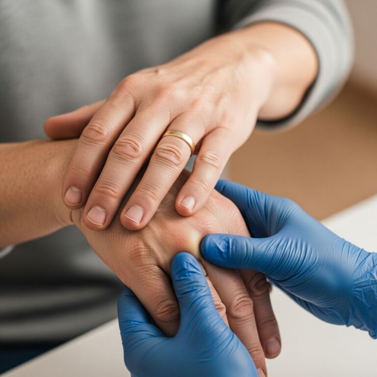
Physical Examination Techniques
After obtaining a comprehensive history, a focused physical examination of the scalp and relevant body areas should be performed. The examination should not be limited to the scalp alone if the patient’s history suggests a systemic condition.
The scalp examination should be conducted systematically, evaluating different regions including the vertex, occipital area, frontal hairline, and postauricular scalp. Visual inspection should assess for erythema, scaling, lichenification, erosions, crusting, and the distribution pattern of lesions. The presence and pattern of hair loss should be noted, including whether alopecia appears diffuse or patchy, and whether hair shafts are broken or completely lost.
Palpation helps determine the texture of scales, presence of nodules or induration, and focal tenderness. The examiner should look for signs of infection such as pustules, purulent drainage, or lymphadenopathy. Documentation of lesion morphology, including size, shape, color, and arrangement, is essential for diagnostic accuracy.
Systematic Examination Approach:
- Inspect scalp under adequate lighting
- Evaluate all scalp regions: vertex, occipital, frontal, temporal, and postauricular areas
- Assess hair quality and presence of hair loss
- Palpate for texture, temperature changes, and tenderness
- Examine adjacent areas (neck, ears, face) for extension of rash
- Check for lymphadenopathy in cervical and occipital nodes
- Assess general skin examination if systemic condition suspected
Diagnostic Testing Methods
While many scalp conditions can be diagnosed clinically based on history and examination, additional diagnostic testing confirms suspected diagnoses and guides treatment.
Trichoscopy (Dermoscopy of the Scalp)
Trichoscopy represents a valuable non-invasive diagnostic tool for evaluating scalp conditions. This technique involves using a handheld dermoscope to magnify and examine the scalp surface and hair structures at high magnification. Trichoscopy provides rapid confirmation of conditions while results of laboratory tests are pending, allowing treatment to commence promptly.
Different scalp conditions demonstrate characteristic trichoscopic findings. For inflammatory conditions such as psoriasis, distinctive findings include red dots, hairpin vessels, and red globular rings. Fungal infections like tinea capitis may demonstrate corkscrew hairs, which are typical of Trichophyton infections, or other characteristic patterns. The trichoscopic examination helps differentiate conditions and confirm clinical diagnoses.
Microscopy and Culture
Microscopic examination of scalp scrapings or plucked hairs provides rapid confirmation of fungal infections. Specimens are prepared by mounting in potassium hydroxide solution and examining under light microscopy for the presence of fungal hyphae and spores. This technique is particularly useful for suspected tinea capitis and other fungal infections.
Fungal culture remains the gold standard for identifying the specific organism causing infection and determining susceptibility to antifungal agents. Culture results guide antifungal selection and help confirm the diagnosis when microscopy is inconclusive.
Skin Biopsy
Skin biopsy is performed when clinical diagnosis remains uncertain after history, examination, and non-invasive testing. A representative lesion is biopsied, processed for histopathological examination, and often includes special stains and cultures when fungal or bacterial infection is suspected. Biopsy may reveal characteristic changes confirming diagnosis and excluding malignancy or other serious conditions.
Common Scalp Conditions and Their Diagnostic Features
Scalp Psoriasis
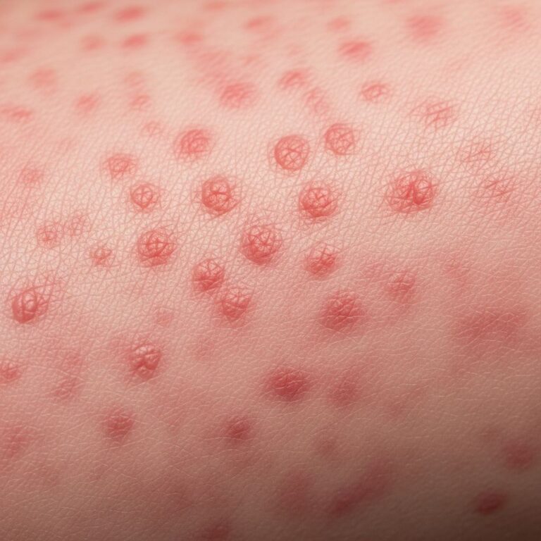
Scalp psoriasis is generally diagnosed clinically based on characteristic features. Key trichoscopic findings include red dots, hairpin vessels, and red globular rings. The condition typically affects the occipital and frontal regions. Skin biopsy may be performed in some cases to confirm diagnosis.
In darker skin phototypes, the presentation may vary significantly from classic descriptions, with purple to dark brown patches and grey or silver scales. This variation can delay diagnosis if not recognized. Assessing psoriasis severity using validated scales such as the Psoriasis Scalp Severity Index (PSSI) and impact on quality of life is important for management and monitoring treatment response.
Tinea Capitis (Scalp Ringworm)
Tinea capitis is a fungal infection involving both scalp skin and hair structures. Symptoms include hair loss, dry scaly areas, redness, and itching. Dermoscopy of the scalp provides rapid confirmation, allowing treatment initiation while awaiting culture results.
Characteristic dermoscopic findings with high predictive value include corkscrew hairs, which are typical of Trichophyton infections. Microscopy of scalp scrapings or plucked hairs can rapidly confirm diagnosis by revealing fungal hyphae and spores when specimens are wet-mounted in potassium hydroxide and examined under light microscopy. Fungal culture identifies the specific organism.
Seborrheic Dermatitis
Seborrheic dermatitis is a common inflammatory skin disease affecting areas rich in sebaceous glands, particularly the scalp. The condition presents with papulosquamous morphology and characteristic distribution. Diagnosis is clinical, based on lesion location, appearance, and behavior.
In infantile cases, seborrheic dermatitis causes cradle cap, presenting as diffuse greasy scaling on the scalp. In children of color, classic cradle cap appearance may not develop; instead, erythema, flaking, and hypopigmentation of affected areas occur. The condition commonly affects the vertex and parietal scalp regions but may extend to facial areas, retroauricular skin, and skin folds.
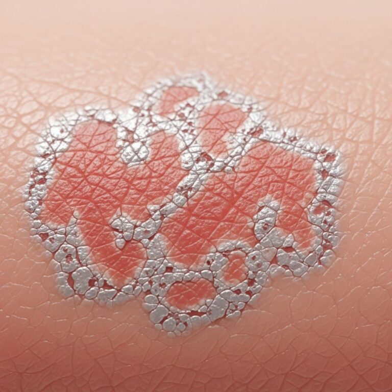
Pityriasis Amiantacea
Pityriasis amiantacea is characterized by excessive scaling with thick silvery or yellowish scales encircling hair shafts. This condition often results from an underlying dermatological disease including seborrheic dermatitis, atopic dermatitis, tinea capitis, head lice, or lichen simplex. Identification of the underlying cause is essential for appropriate treatment.
Skin and hair samples for mycology and bacterial culture help identify infectious etiologies. Skin biopsy is rarely necessary. Treatment depends on addressing the specific underlying disease.
Differential Diagnosis Considerations
The differential diagnosis list for scalp rashes is extensive, encompassing any condition presenting with patchy alopecia, inflammation, or scaling. Systematic evaluation helps distinguish among these possibilities.
Distinctions between similar conditions are important. Psoriasis tends to affect the occipital and frontal regions with mostly non-pruritic presentation, whereas seborrheic dermatitis affects the vertex and parietal regions. Contact eczema may result from shampoo, hair dye, or other product allergies. Tinea capitis presents with broken hairs or characteristic “black dots” (arthroconidia-filled follicles) and is uncommon in adults. Impetigo presents with yellow, honey-colored crusting and suggests bacterial infection.
Key Differential Diagnoses:
- Psoriasis — non-pruritic, occipital/frontal distribution
- Seborrheic dermatitis — vertex/parietal distribution, greasy appearance
- Tinea capitis — fungal infection, broken hairs, uncommon in adults
- Contact dermatitis — related to product exposure
- Atopic dermatitis — pruritic, history of atopy
- Head lice — visible nits, intense pruritus
- Lichen planus — violet papules, linear arrangement
- Folliculitis — pustules, hair follicle involvement
- Darier disease — yellowish-brown papules in seborrheic distribution
Diagnostic Testing Summary Table
| Test | Indications | Advantages |
|---|---|---|
| Trichoscopy | Suspected tinea capitis, psoriasis, inflammatory conditions | Non-invasive, rapid, guides treatment initiation |
| Microscopy (KOH) | Suspected fungal infection | Rapid results, identifies fungal elements |
| Fungal Culture | Confirmed or suspected tinea capitis | Identifies organism, guides therapy selection |
| Bacterial Culture | Suspected bacterial infection or folliculitis | Identifies pathogen, determines susceptibility |
| Skin Biopsy | Uncertain diagnosis, chronic conditions, suspected malignancy | Definitive histological diagnosis, special stains available |
Special Considerations in Diagnosis
Certain populations require special diagnostic consideration. In patients with darker skin phototypes, scalp conditions may present with different morphologies than classic descriptions. Psoriasis and seborrheic dermatitis may appear as purple to dark brown patches rather than the classic red or salmon coloration. Recognition of these variations prevents delayed or missed diagnosis.
Hair texture and styling practices also influence presentation. Conditions associated with tight hairstyling may present with localized inflammation or traction alopecia. When formulating treatment plans, compatibility with patient hair care practices and cultural preferences is essential for adherence and effectiveness.
When to Refer to a Specialist
Referral to a dermatologist is appropriate when scalp rashes persist despite appropriate treatment, diagnosis remains uncertain after initial evaluation, systemic involvement is suspected, or rare conditions are encountered. Dermatologists can provide specialized diagnostic testing including trichoscopy and dermoscopic interpretation, perform skin biopsy when indicated, and manage complex or refractory conditions.
Frequently Asked Questions
Q: How can I differentiate between scalp psoriasis and seborrheic dermatitis?
A: Psoriasis typically presents as non-pruritic lesions affecting the occipital and frontal regions, while seborrheic dermatitis tends to involve the vertex and parietal regions and may appear greasy. Trichoscopy showing red dots, hairpin vessels, and red globular rings suggests psoriasis. Clinical presentation and distribution pattern are usually diagnostic, though biopsy can confirm if needed.
Q: Is trichoscopy painful or invasive?
A: No, trichoscopy is a non-invasive procedure using a handheld dermoscope to magnify scalp structures. It causes no pain or tissue damage and can be performed in an office setting without anesthesia, making it an excellent rapid diagnostic tool.
Q: What should I expect during a scalp biopsy?
A: A biopsy involves removing a small representative lesion under local anesthesia. The tissue is examined microscopically for diagnostic characteristics. Discomfort is minimal with proper anesthesia, and scarring is usually minimal when performed by experienced clinicians on hidden scalp areas.
Q: How long do fungal culture results take?
A: Fungal cultures typically require 2-4 weeks for definitive organism identification and susceptibility testing. This is why trichoscopy and KOH microscopy are valuable for initiating treatment while awaiting culture confirmation.
Q: Can scalp rashes be diagnosed without visiting a doctor?
A: While self-diagnosis is tempting, professional evaluation is recommended for accurate diagnosis and appropriate treatment. Many scalp conditions require clinical assessment, and some require diagnostic testing for confirmation. Misdiagnosis can lead to ineffective or inappropriate treatment, prolonging symptoms.
Q: Why is my scalp rash misdiagnosed in dermatology clinics?
A: Scalp conditions may be missed or misdiagnosed due to overlapping presentations, subtle variations in darker skin types, incomplete examination of all scalp regions, or failure to perform appropriate diagnostic testing. Thorough history, systematic examination, and appropriate testing minimize diagnostic errors.
References
- Diagnosis of Scalp Rashes — DermNet. Last updated February 2016. https://dermnetnz.org/topics/diagnosis-of-scalp-rashes
- Scalp Psoriasis: A Complete Overview — DermNet. https://dermnetnz.org/topics/scalp-psoriasis
- Tinea Capitis — DermNet. https://dermnetnz.org/topics/tinea-capitis
- Seborrheic Dermatitis: Causes and Treatment — DermNet. https://dermnetnz.org/topics/seborrhoeic-dermatitis
- Seborrheic Dermatitis — StatPearls, National Center for Biotechnology Information. https://www.ncbi.nlm.nih.gov/books/NBK551707/
- Pityriasis Amiantacea — DermNet. https://dermnetnz.org/topics/pityriasis-amiantacea
Similar Articles


Read full bio of medha deb