Diffuse Dermal Angiomatosis of the Breast
Rare vascular skin disorder causing severe breast pain, violaceous plaques, and ulcerations linked to tissue hypoxia.

Diffuse dermal angiomatosis of the breast (DDAB) is a rare benign vascular proliferation disorder characterized by severe breast pain and distinctive skin lesions.
What is diffuse dermal angiomatosis of the breast?
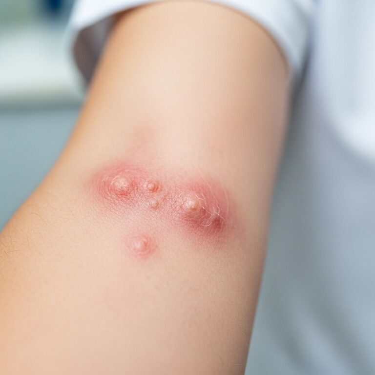
Diffuse dermal angiomatosis (DDA) represents a rare variant of reactive cutaneous angioendotheliomatosis (RAE), featuring excessive endothelial cell proliferation in the reticular dermis. When affecting the breasts, known as DDAB, it manifests as tender, violaceous to erythematous plaques often with central ulcerations and reticular patterns. This condition typically arises in middle-aged women with pendulous breasts or vascular risk factors, leading to tissue hypoxia that triggers neovascularization.
Clinically, DDAB presents with excruciating, intractable pain disproportionate to visible changes, alongside benign-appearing purpuric papules and indurated plaques that progressively enlarge. Lesions commonly involve the dependent portions of large breasts, exacerbated by gravitational ischemia.
Who gets diffuse dermal angiomatosis of the breast?
DDAB predominantly affects middle-aged to older women, often those with large, pendulous breasts (macromastia). A case series documented eight patients averaging 58 years old, all female with bilateral involvement in most cases. Risk factors mirror systemic vascular compromise:
- Atherosclerosis and peripheral vascular disease
- Smoking history
- Obesity and diabetes
- Hyperlipidemia
- Macromastia causing mechanical compression
These factors promote chronic tissue hypoxia, stimulating compensatory angiogenesis.
Clinical features
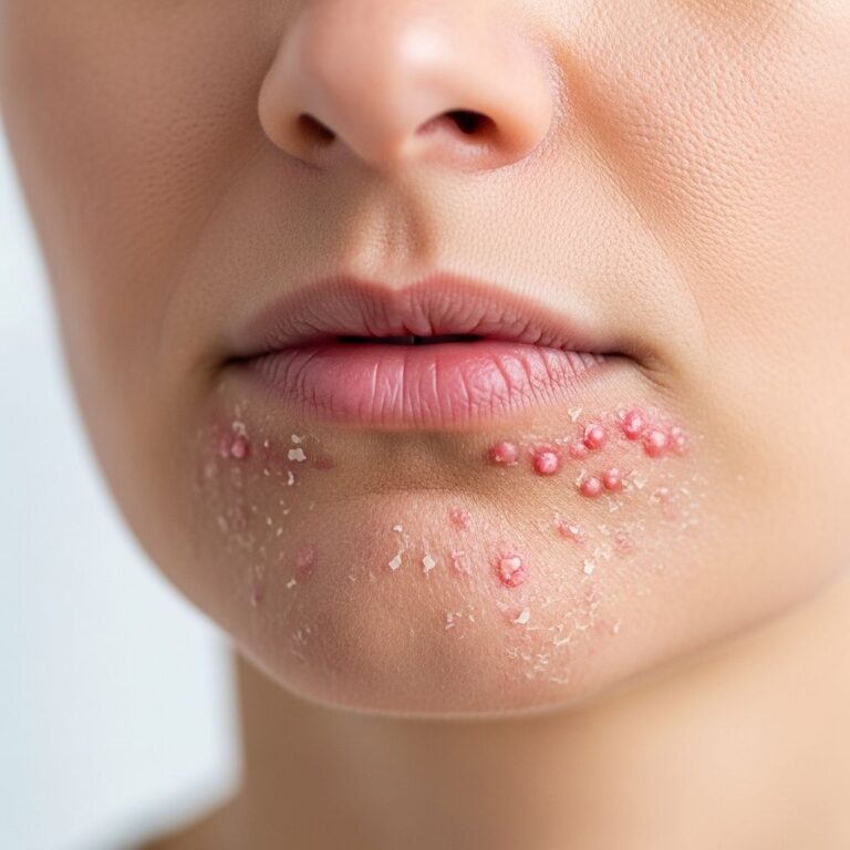
Patients report unbearable breast pain lasting weeks to months, often starting unilaterally before becoming bilateral. Skin findings include:
- Reticular violaceous erythematous plaques with livedo-like patterns
- Central ulcerations and crusting
- Indurated, tender purpuric papules
- Surrounding telangiectasias and visible vessels
Lesions favor inframammary folds and inferior breast surfaces due to pressure and poor perfusion. Pain resists standard analgesics, prompting extensive evaluation.
Diagnosis
Diagnosis requires clinicopathologic correlation with punch biopsy essential for confirmation. Key diagnostic steps:
- Clinical recognition of characteristic reticular plaques with ulceration
- 4-6mm punch biopsy of lesional and perilesional skin
- Immunohistochemistry (CD31, CD34, ERG positive endothelial markers)
- Exclusion of systemic vascular occlusion via MRI/MRA/MRV
Histology reveals diffuse dermal capillary proliferation with plump endothelial cells lining irregular vascular channels interspersed among collagen bundles. Vessel lumens may appear obliterated, with occasional erythrocytes but absent thrombi or atypia distinguishing from malignancy.
Differential diagnosis
| Condition | Key Distinguishing Features |
|---|---|
| Kaposi sarcoma | HHV-8 positive, spindle cells, slit-like spaces |
| Angiosarcoma | Atypical mitoses, deep infiltration, anastomosing channels |
| Acroangiodermatitis | Pseudo-Kaposi, legs, venous insufficiency |
| Reactive angioendotheliomatosis | Intraluminal proliferation vs. interstitial in DDA |
| Inflammatory breast carcinoma | Pagetoid changes, malignant cells on biopsy |
Investigations
Workup excludes mimics and identifies hypoxia drivers:
- Imaging: Breast ultrasound/MRI rules out malignancy; MRA/MRV assesses vasculature
- Labs: Lipid panel, HbA1c, CBC for comorbidities
- Vascular studies: ABI, Doppler for peripheral disease
- Biopsy: H&E plus IHC (CD31+, ERG+, pan-cytokeratin negative)
Management
No standardized guidelines exist; treatment targets underlying hypoxia and vascular proliferation. Multimodal approach yields best outcomes:
Conservative measures
- Smoking cessation
- Weight loss
- Breast reduction surgery for macromastia
- Compression garments
Medical therapy
- Isotretinoin: 0.5-1mg/kg/day led to rapid pain resolution and lesion regression in reported cases
- Antiplatelets (aspirin)
- Statins for atherosclerosis
- Topical steroids, wound care for ulcers
Surgical interventions
- Revascularization for occlusive disease
- Mastectomy rarely for refractory cases
Isotretinoin success likely stems from antiangiogenic effects and sebaceous gland reduction improving local perfusion.
Prognosis and complications
DDAB follows a chronic, relapsing course without intervention, with persistent pain and ulceration risking infection. Addressing risk factors achieves remission in most; recurrence ties to ongoing hypoxia. Complications include secondary bacterial infection and scarring.
Frequently Asked Questions
Is diffuse dermal angiomatosis of the breast cancerous?
No, DDAB is a benign reactive process despite alarming appearance. Biopsy excludes angiosarcoma via absent atypia.
What causes the severe pain in DDAB?
Pain arises from ischemic tissue and nerve compression by proliferating vessels, amplified by breast weight.
How is DDAB diagnosed?
Definitive diagnosis requires skin biopsy showing dermal endothelial proliferation confirmed by CD31/ERG stains.
Does DDAB only affect the breasts?
While classic on legs, breast involvement occurs with macromastia; systemic cases reported.
Can DDAB be cured?
Treatment of underlying vascular disease and/or isotretinoin achieves prolonged remission in most patients.
Conclusion
Diffuse dermal angiomatosis of the breast remains underrecognized yet treatable when identified early. Clinicians must maintain high suspicion for this entity in women with refractory breast pain and reticular violaceous plaques, pursuing biopsy to guide hypoxia-directed therapy.
References
- A cause of unbearably painful breast, diffuse dermal angiomatosis — PMC. 2013-11-20. https://pmc.ncbi.nlm.nih.gov/articles/PMC3864042/
- A cause of unbearably painful breast, diffuse dermal angiomatosis — AME Groups. 2013. https://gs.amegroups.org/article/view/1009/html
- Diffuse dermal angiomatosis of the breast — PMC. 2020-04-01. https://pmc.ncbi.nlm.nih.gov/articles/PMC7155956/
- Diffuse Dermal Angiomatosis of the Breast — JAMA Dermatology. 2000. https://jamanetwork.com/journals/jamadermatology/fullarticle/403400
- Diffuse Dermal Angiomatosis of the Breasts: A Case Series of 8 — DermSquared. N/A. https://skin.dermsquared.com/skin/article/view/1191
- Diffuse Dermal Angiomatosis — The Hospitalist. N/A. https://blogs.the-hospitalist.org/content/diffuse-dermal-angiomatosis
Similar Articles


Read full bio of medha deb