Digital Myxoid Cyst: Causes, Symptoms, and Treatment
Complete guide to digital myxoid cysts: causes, identification, and management options.

What is a Digital Myxoid Cyst?
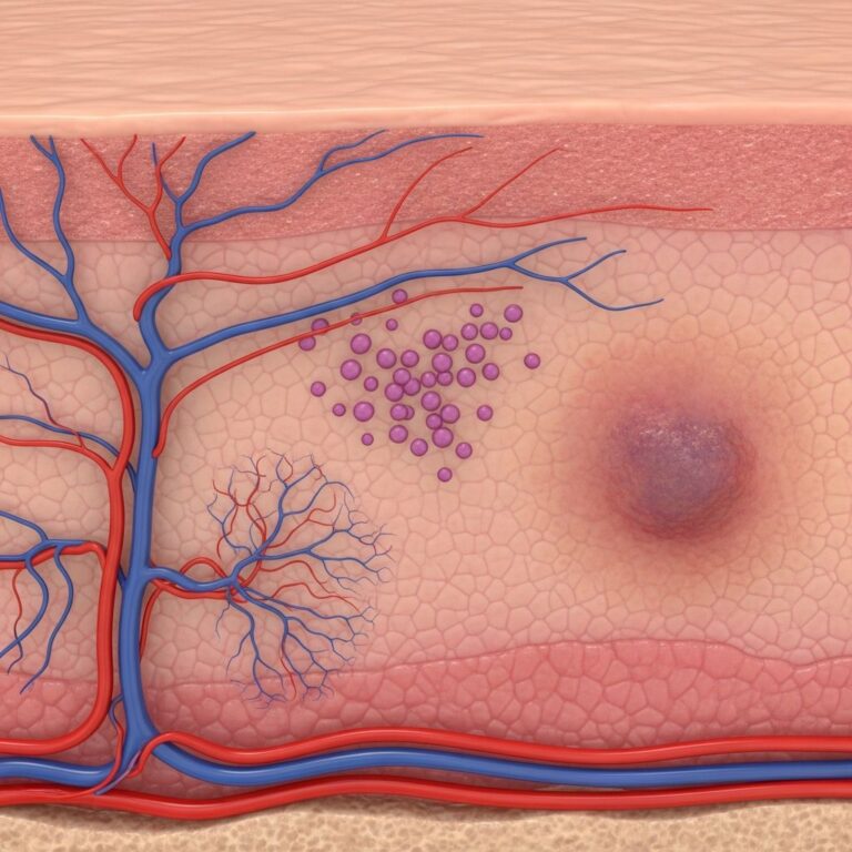
A digital myxoid cyst, also known as a digital mucous cyst or mucous pseudocyst, is a small, benign (non-cancerous) swelling that typically develops on the fingers or, less commonly, on the toes. This condition represents one of the most frequent cystic lesions affecting the distal digits, occurring most commonly in adults over 40 years of age. The cyst is often connected to the joint lining at the end of the finger or toe and usually appears as a smooth, shiny papule with a distinctive appearance.
These lesions are characterized by their semi-translucent quality and smooth, glistening surface that gives them a pearl-like appearance. The cysts typically range in size from 5 millimeters to 1 centimeter across, roughly equivalent to the size of a pencil eraser. Most people develop only a single cyst, though some individuals may develop multiple cysts on the same or different fingers.
Physical Characteristics and Appearance
Digital myxoid cysts present with distinctive visual and tactile features that aid in identification:
- Color: Usually skin-colored or translucent with possible reddish or bluish tinge, often resembling a small pearl
- Surface texture: Smooth and shiny with a firm or fluid-filled consistency
- Size: Typically 5 mm to 1 cm in diameter, comparable to a pencil eraser
- Shape: Round or oval bumps that develop beneath the skin surface
- Location: Most commonly found near the nail on the index, middle, or ring finger of the dominant hand, with attachment to the joint by a thin stalk
- Contents: Jelly-like, sticky fluid that may ooze from the cyst, sometimes blood-stained
The characteristic groove in the adjacent fingernail serves as an important diagnostic feature, as the cyst’s pressure on the nailbed beneath the skin surface can create a distinctive dent or groove that extends down the length of the nail. This nail involvement may lead to slight depressions, color changes, nail splitting, or in severe cases, nail loss.
Causes and Risk Factors
The exact etiology of digital myxoid cysts remains unclear, though researchers have identified two primary mechanisms of formation:
Joint Degeneration Theory
One explanation involves degeneration of synovial tissue surrounding the finger or toe joint, typically associated with osteoarthritis and other degenerative joint diseases. In this variation, a small bony growth formed from degenerating joint cartilage, known as an osteophyte, may be involved. This type of cyst functions as a type of ganglion, with a stalk that tracks back to the joint.
Metabolic Derangement Theory
The second mechanism suggests that fibroblast cells in the connective tissue produce excessive amounts of mucin, an ingredient of mucus. This variation represents a form of focal mucinosis, characterized by abnormal deposits of mucopolysaccharides in the skin, and does not necessarily involve joint degeneration.
Risk Factors
Several factors increase susceptibility to developing digital myxoid cysts:
- Age: Most frequently occur in people in their sixties, though they can develop at any age
- Gender: Affect females approximately three times as often as males
- Osteoarthritis: Strong association with underlying osteoarthritis, with 64% to 93% of people with osteoarthritis developing these cysts
- Trauma history: In some cases, there may be a history of trauma to the affected finger or toe
- Repetitive motion: Some individuals develop cysts due to excessive repetitive finger motion
Symptoms and Clinical Presentation
Digital myxoid cysts typically present with minimal symptoms, though clinical manifestations can vary:
Primary Symptoms
- Slow growth: Cysts develop gradually over months
- Pain characteristics: Usually painless, though they may become tender when knocked or during joint movement
- Fluid discharge: Clear, straw-colored, or blood-stained sticky fluid may occasionally leak from the cyst
- Joint symptoms: Arthritis pain, stiffness, and deformity of the adjacent joint may occur
- Nail changes: Grooves extending down the nail length, slight depressions, color changes, splitting, or nail loss when the cyst affects the nail root
- Skin changes: Possible skin thinning or redness over the bump
Subungual Involvement
When a myxoid cyst grows under the nail or involves the nail root, the condition can become painful. This type of cyst may cause significant nail deformity and functional impairment, warranting consideration of treatment options.
Diagnosis
Medical professionals typically diagnose digital myxoid cysts through straightforward clinical assessment:
- Physical examination: Visual inspection and palpation of the small lump overlying the end joint of the finger or toe
- Visual characteristics: Recognition of the distinctive smooth, shiny, semi-translucent papule
- Nail involvement: Observation of any groove in the adjacent fingernail
- Clinical history: Questions about the cyst’s duration, growth rate, symptoms, and any recent trauma or infection
- Fluid assessment: Occasional discharge of clear, slightly straw-colored, or blood-stained fluid aids diagnosis
The diagnosis is usually straightforward based on clinical presentation, and additional diagnostic imaging or laboratory tests are rarely necessary for typical presentations.
Treatment Options
Management of digital myxoid cysts depends on symptom severity and patient preference:
Conservative Management
Most digital myxoid cysts do not require active treatment. Many individuals find that conservative approaches suffice:
- Observation: Many cysts remain stable or may spontaneously resolve over time
- Repeated pressure: Firmly pressing on the cyst repeatedly for several weeks may help reduce its size in some cases, though effectiveness varies
- Joint care: Managing underlying osteoarthritis symptoms may help prevent progression
Surgical Intervention
When treatment is desired, surgical removal represents the most effective method:
- Complete excision: Surgical removal of the entire cyst structure provides optimal results
- Joint cleanup: In some cases, removal of bony spurs or joint surface irregularities may reduce recurrence
- Stalk removal: Complete removal of the stalk connecting the cyst to the joint is crucial for minimizing recurrence
Important Considerations
Before pursuing treatment, patients should understand important limitations:
- Self-drainage: Attempting to drain or puncture the cyst yourself should be avoided, as this can cause infection
- Recurrence rates: Recurrence rates range from 14% to 70% depending on the treatment method employed
- Side effects: Treatment options often come with side effects including pain and scarring
- Risk-benefit analysis: The potential benefits of treatment must be weighed against the risk of recurrence and possible complications
When to Seek Medical Attention
Although most digital myxoid cysts are benign and self-limiting, certain situations warrant professional evaluation:
- Sudden enlargement of the cyst accompanied by redness and warmth, suggesting infection
- Significant pain or functional impairment affecting daily activities or work
- Cosmetic concerns that bother the patient
- Significant nail deformity or loss affecting appearance or function
- Signs of infection such as increased warmth, redness, swelling, or purulent drainage
- Uncertainty about diagnosis or desire for professional assessment
Distinguishing from Other Conditions
Digital myxoid cysts differ from similar conditions in important ways. Ganglion cysts, for comparison, are generally caused by joint or tendon irritation rather than joint wear and arthritis. While myxoid cysts typically affect the distal fingers near the nail, ganglion cysts more commonly occur on the wrist or hand. Understanding these distinctions helps ensure appropriate diagnosis and management.
Frequently Asked Questions
Q: Are digital myxoid cysts cancerous?
A: No, digital myxoid cysts are benign (non-cancerous) growths. They pose no risk of malignant transformation and are not life-threatening.
Q: Can I prevent digital myxoid cysts from developing?
A: Since the exact cause remains unknown, complete prevention is not possible. However, managing osteoarthritis and avoiding repetitive trauma to the fingers may reduce risk in some individuals.
Q: Will my digital myxoid cyst go away on its own?
A: Some cysts may remain stable or spontaneously resolve, though this varies by individual. Most do not require treatment and can be safely monitored over time.
Q: What happens if I ignore my digital myxoid cyst?
A: In most cases, leaving a cyst untreated poses no serious health risk. However, if nail involvement causes significant deformity or if the cyst becomes infected, professional evaluation is recommended.
Q: How long does it take a digital myxoid cyst to develop?
A: Digital myxoid cysts grow slowly over months. The rate of growth varies considerably among individuals.
Q: Are there any home remedies for digital myxoid cysts?
A: Firmly pressing on the cyst repeatedly for several weeks may help reduce size in some cases. However, attempting to drain or puncture the cyst yourself should be avoided due to infection risk.
Q: What is the success rate of surgical removal?
A: While surgical removal is effective, recurrence rates range from 14% to 70% depending on the surgical technique and completeness of removal, particularly regarding stalk removal.
References
- Myxoid cyst (digital mucous cyst) — Primary Care Dermatology Society. 2024. https://www.pcds.org.uk/patient-info-leaflets/myxoid-cyst-digital-mucous-cyst
- Digital Myxoid Cyst: Causes, Symptoms, Pictures, and Treatment — Healthline Media. 2024. https://www.healthline.com/health/myxoid-cyst
- Digital Myxoid cyst — Skin Health Info. 2019. https://www.skinhealthinfo.org.uk/wp-content/uploads/2018/11/Digital-myxoid-cyst-PIL-July-2019.pdf
- Myxoid Cyst (Digital Mucous Cyst): Causes & Treatment — Cleveland Clinic. 2024. https://my.clevelandclinic.org/health/diseases/23456-myxoid-cyst
- Digital mucous (myxoid) cyst — DermNet. 2024. https://dermnetnz.org/topics/digital-myxoid-pseudocyst
- Office Management of Digital Mucous Cysts — American Family Physician, 2001. https://www.aafp.org/pubs/afp/issues/2001/1215/p1987.html
- What Are Myxoid Cysts? How They Compare to Ganglion Cysts — Banner Health. 2024. https://www.bannerhealth.com/healthcareblog/teach-me/what-are-myxoid-cysts
Similar Articles


Read full bio of Sneha Tete