Digital Myxoid Pseudocyst: What You Need To Know
Understanding digital myxoid pseudocysts: benign finger cysts linked to joint degeneration, nail changes, and effective treatments.

A
digital myxoid pseudocyst
, also known as a digital mucous cyst, myxoid cyst, or digital ganglion cyst, is a benign, fluid-filled lesion that appears as a shiny papule on the distal end of a finger or toe, close to the nail. Despite its name, it is a pseudocyst lacking a true epithelial lining, filled instead with thick, gelatinous mucin rich in hyaluronic acid.What is a digital myxoid pseudocyst?
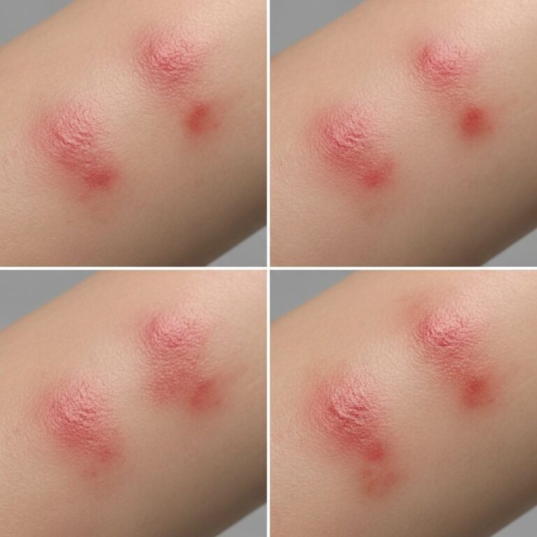
Digital myxoid pseudocysts are common benign tumours of the digits, representing up to 12% of nail unit tumours in some series. They typically manifest as solitary, dome-shaped, translucent or skin-coloured papules or nodules, ranging from a few millimetres to 1 cm in size, located on the dorsal aspect near the distal interphalangeal (DIP) joint or proximal nail fold. The lesion arises from degeneration in the connective tissue overlying the DIP joint, often associated with osteoarthritis.
Clinically, these pseudocysts have a smooth, shiny surface and may appear semi-translucent, allowing light to pass through (transillumination test positive). They contain jelly-like, sticky fluid that can sometimes be expressed, occasionally tinged with blood. Patients usually present due to cosmetic concerns, nail deformity such as a longitudinal groove, or mild discomfort rather than severe pain. Spontaneous rupture can occur, leading to crusting, ulceration, or secondary infection.
Who gets digital myxoid pseudocysts?
Digital myxoid pseudocysts predominantly affect middle-aged to elderly adults, with a peak incidence in those over 50 years. Women are affected more frequently than men, possibly due to hormonal or occupational factors. They are almost exclusively located on the fingers, particularly the index, middle, and ring fingers, though toes can be involved. Association with underlying DIP joint osteoarthritis is common, especially in patients with Heberden nodes. Trauma or repetitive stress to the digits may precipitate formation in susceptible individuals.
What causes digital myxoid pseudocysts?
The exact aetiology involves two main pathogenic variants:
- Ganglion-type: An outpouching or extension of the synovial lining from the DIP joint, often with a fibrous stalk connecting the pseudocyst to the joint space. This is linked to osteoarthritis, where joint degeneration allows synovial fluid extravasation.
- Myxomatous-type: Focal mucoid degeneration of dermal collagen, with fibroblasts producing excessive hyaluronic acid and mucopolysaccharides, independent of the joint.
Additional theories include mucin deposition from chronic trauma or stress. Histologically, both types show myxoid degeneration without neoplastic cells. Osteophytes at the DIP joint are frequently observed, displacing the extensor tendon and promoting lateral cyst formation.
What are the clinical features of digital myxoid pseudocysts?
Lesions are typically solitary, round to oval, and located within 1 cm of the nail base, often just off the midline (radial or ulnar to the extensor tendon). Key features include:
- Dome-shaped, translucent nodule with shiny surface.
- Size: 2–10 mm diameter.
- May cause a longitudinal nail groove (Type B lesion) due to compression of the germinal matrix; groove can vary in depth with cyst fluctuation (“waves sign” on dermoscopy).
- Rarely painful, but associated joint stiffness or arthritis symptoms possible.
- Spontaneous rupture releases viscous mucin, followed by crust or ulcer.
Dermoscopy reveals structureless bluish areas, vascular patterns (arborizing, dotted, linear vessels), and red-purple lacunae. Classification by location (de Berker et al.): Type A (dorsal skin between DIP and proximal nail fold), Type B (within proximal nail fold, nail groove), Type C (subungual, rare).
Diagnosis
Diagnosis is primarily clinical, based on characteristic location, appearance, and demographics. Transillumination confirms fluid content. Dermoscopy aids differentiation from warts, basal cell carcinoma, or melanoma. Ultrasound may demonstrate connection to the DIP joint or multilocular fluid collections. Histology, if biopsied, shows no epithelial lining, dense collagen wall, fibroblasts, and Alcian blue-positive mucin pools. No atypia present, confirming benign nature.
What is the differential diagnosis for digital myxoid pseudocysts?
| Condition | Key Distinguishing Features |
|---|---|
| Verruca vulgaris | Hyperkeratotic surface, thrombosed capillaries on dermoscopy, HPV association. |
| Onychomatricoma | Nail plate thickening, filamentous projections on imaging. |
| Basal cell carcinoma | Pearly border, telangiectasia, rolled edges. |
| Gouty tophus | White chalky material, associated hyperuricaemia. |
| Pyogenic granuloma | Collarette scale, rapid growth, bleeding tendency. |
| Melanoma (nail apparatus) | Irregular pigmentation, Hutchinson sign. |
Radiographs may reveal DIP osteoarthritis or osteophytes to support diagnosis.
Pathology
Histopathology reveals a multiloculated gelatinous nodule without true cyst lining. The wall comprises compressed collagen, fibroblasts, and capillary proliferation blending into dermis. Cavity filled with hyaluronic acid-rich mucin (Alcian blue positive). Peripheral degenerative collagen fragmentation common. Stalk, if present, shows fibrous connection to joint or tendon sheath. Overlying epidermis thinned. Special stains confirm glycosaminoglycans; no IHC typically needed.
Treatment of digital myxoid pseudocysts
Treatment is indicated for cosmetic reasons, nail deformity, or recurrent rupture/infection. Options include:
- Observation: For asymptomatic small lesions.
- Aspiration ± steroid injection: Simple office procedure; recurrence high (50–70%).
- Sclerotherapy: Injection of sclerosant (e.g., sodium tetradecyl sulfate).
- Excision: Surgical removal of cyst and stalk, with joint capsule ligation if connected; lowest recurrence (5–10%).
- Laser therapy or cryotherapy: For superficial lesions.
Post-treatment, nail groove may persist but often improves. Recurrence risk higher without addressing underlying osteoarthritis.
Prevention
Preventive measures are limited, but managing osteoarthritis (e.g., hand therapy, anti-inflammatories) and avoiding digit trauma may help. Early intervention prevents complications like infection.
References
References
- Digital mucous (myxoid) cyst — DermNet NZ. 2023. https://dermnetnz.org/topics/digital-myxoid-pseudocyst
- Mucous Pseudocyst (Digital Myxoid Cyst) — DermaCompass. Accessed 2026. https://www.dermacompass.net/en/diseases/mucous-pseudocyst-digital-myxoid-cyst
- The “Waves Sign” in Digital Myxoid Pseudocyst — PMC – NIH. 2024-05-29. https://pmc.ncbi.nlm.nih.gov/articles/PMC11147518/
- Digital Mucous Cyst — StatPearls – NCBI Bookshelf. 2023. https://www.ncbi.nlm.nih.gov/books/NBK559092/
- Myxoid cyst (digital mucous cyst) — Primary Care Dermatology Society (PCDS). Accessed 2026. https://www.pcds.org.uk/patient-info-leaflets/myxoid-cyst-digital-mucous-cyst
Frequently asked questions
What is a digital myxoid pseudocyst?
A benign, translucent, jelly-filled lump near the nail on fingers or toes, arising from joint or connective tissue degeneration.
Is a digital myxoid pseudocyst painful?
Usually not; discomfort arises from associated arthritis or rupture.
Do digital myxoid pseudocysts go away on their own?
They may persist or spontaneously rupture but often recur without treatment.
How is it treated?
Options range from observation to surgical excision; surgery offers lowest recurrence.
Can it affect the nail?
Yes, often causing a longitudinal groove due to proximal nail fold compression.
Is it cancerous?
No, entirely benign with no malignant potential.
Similar Articles


Read full bio of medha deb