Disorders Of Keratinisation: Diagnosis, Genetics, And Treatment
Comprehensive guide to disorders of keratinisation causing scaly rashes, from ichthyosis to Darier disease.

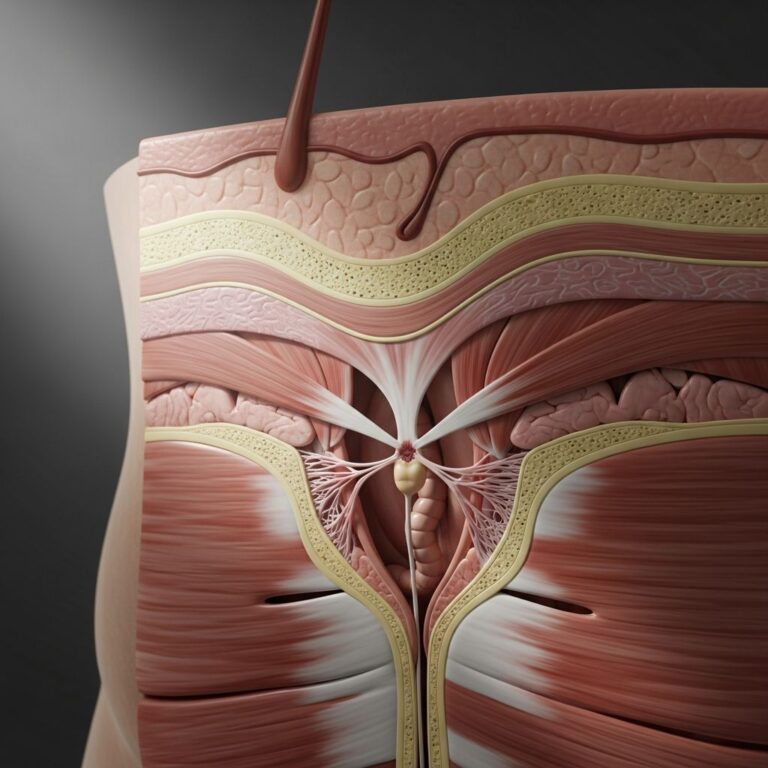
Disorders of keratinisation, also known as disorders of cornification, encompass a diverse group of genetic and acquired conditions characterised by abnormal differentiation and desquamation of keratinocytes, leading to excessively dry, scaly skin. These conditions affect the epidermis, resulting in persistent scaling, hyperkeratosis, and sometimes associated inflammation or blistering. Genetic mutations in keratin proteins, filaggrin, transglutaminase, and other structural elements disrupt the normal process of cornification, where keratinocytes transform into the protective stratum corneum.
Understanding these disorders is crucial for dermatologists, as they present with distinctive clinical patterns that guide diagnosis and management. This article reviews key disorders, their histopathology, molecular genetics, and therapeutic approaches, drawing from peer-reviewed literature and clinical guidelines.
Ichthyosis
Ichthyosis refers to a spectrum of disorders marked by generalised dry skin and prominent scaling due to defective epidermal barrier function. The term derives from the Greek ‘ichthys’ meaning fish, reflecting the fish-like scales. Genetic ichthyoses arise from mutations in genes encoding epidermal structural proteins, while acquired forms associate with malignancy or systemic disease.
Common genetic subtypes include:
- Ichthyosis vulgaris (autosomal dominant): Most prevalent, affecting 1 in 250 individuals. Caused by mutations in the filaggrin gene (FLG), leading to reduced natural moisturising factor. Features fine, white scales on extensor limbs, sparing flexures; often begins in childhood and worsens in winter.
- Autosomal recessive lamellar ichthyosis: Severe form due to transglutaminase-1 (TGM1) mutations. Infants born as collodion babies (membrane-covered), developing large, dark plate-like scales over trunk and limbs, ectropion, and palmoplantar keratoderma. Associated with frequent infections.
- X-linked recessive ichthyosis: Affects males, caused by steroid sulfatase (STS) deficiency. Dirty brown scales on neck, trunk, and extremities; corneal opacities in 20%. Diagnosed by elevated cholesterol sulfate levels.
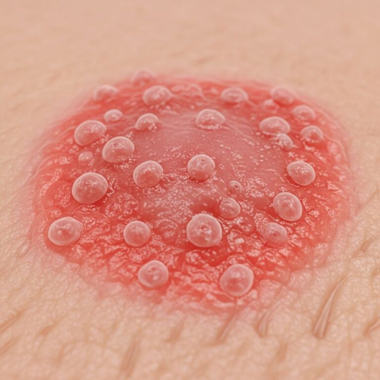
- Bullous ichthyosiform erythroderma (epidermolytic ichthyosis): Mutations in keratin 1 (KRT1) or 10 (KRT10). Presents at birth with erythroderma and fragility blistering, evolving to verrucous hyperkeratosis. Prone to secondary bacterial infections.
Acquired ichthyosis signals underlying pathology like lymphoma, cachexia, or drug reactions (e.g., kava consumption). It mimics genetic forms but onset is later in life.
Management of Ichthyosis
Treatment focuses on hydration, keratolysis, and anti-inflammation. Daily emollients with urea (10-20%) or propylene glycol soften scales. Topical retinoids (tazarotene) promote desquamation but irritate. Oral retinoids (acitretin) for severe cases, monitored for hyperlipidaemia and teratogenicity. Antibiotics for infections.
Palmoplantar Keratodermas
Palmoplantar keratodermas (PPK) feature localised hyperkeratosis of palms and soles, classified as diffuse, focal, or striate. They may be isolated or syndromic, linked to keratin mutations (e.g., KRT1, KRT9) causing mechanical instability.
| Type | Genetics | Clinical Features | Management |
|---|---|---|---|
| Diffuse PPK (epidermolytic) | KRT1/KRT10 | Yellow-brown hyperkeratosis, blistering | Retinoids, salicylic acid |
| Focal non-epidermolytic | KRT9 | Corn-like thickenings on pressure sites | Keratolytics, emollients |
| Mal de Meleda | SLURP1 | Transgrediens spread, autoamputation | Acitretin |
| Syndromic (e.g., Vohwinkel) | GJB2 | PPK + starfish keratosis, deafness | Symptom control |
PPK increases infection risk and impairs function; aggressive debridement and topical therapies are essential.
Keratosis Pilaris
Keratosis pilaris (KP) is a common follicular hyperkeratosis presenting as rough, follicular papules with surrounding erythema on upper arms, thighs, and cheeks. It reflects plugged follicles with absent or reduced filaggrin, akin to ichthyosis vulgaris. Often asymptomatic but cosmetically distressing; persists lifelong with flares.
Treatment yields modest improvement: urea/lactic acid emollients hydrate, topical retinoids (adapalene) normalise keratinisation, but irritation limits use. Laser therapy for refractory cases.
Pityriasis Rubra Pilaris
Pityriasis rubra pilaris (PRP) comprises rare inflammatory disorders with orange-red scaly plaques, follicular papules, and palmoplantar keratoderma. Classic adult type features ‘islands of sparing’; juvenile forms progress to erythroderma. Histology shows alternating ortho- and parakeratosis. Aetiology unknown; possible autoimmune.
Therapy: potent topical steroids, methotrexate (first-line systemic), acitretin, or biologics (e.g., ustekinumab) for refractory disease. Spontaneous remission in 50-80% over 3 years.
Darier Disease
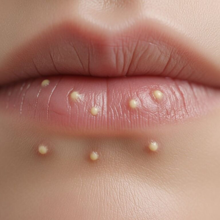
Darier disease (keratosis follicularis, DD) is an autosomal dominant genodermatosis from ATP2A2 mutations impairing desmosomal function. Greasy, crusted yellow-brown papules coalesce on seborrhoeic areas (scalp, chest, flexures); nail dystrophy and palmoplantar pits common. Oral cobblestone papules on palate. Worsened by heat, UV, infection.
Management: sun protection, topical retinoids/steroids, oral retinoids (acitretin 0.5-1mg/kg). Antibiotics for superinfection; HSV prophylaxis.
Lichen Planus and Variants
Idiopathic lichen planus (LP) shows polygonal violaceous papules with Wickham striae, Wickham striae on mucosa. Follicular LP (lichen planopilaris) causes scarring alopecia with perifollicular scaling. Histology: lichenoid infiltrate, basal vacuolisation, Civatte bodies.
Lichen sclerosus (LS), distinct from LP, affects anogenital skin with white sclerotic plaques, atrophy, and purpura. Autoimmune; topical ultrapotent steroids (clobetasol) mainstay, with dermatologist oversight.
Other Keratinisation Disorders
- Olmsted syndrome: Mutilating PPK with periorificial keratoses; TRPV3 mutations.
- Pachyonychia congenita: KRT6A/B/16/17 mutations; painful PPK, oral leukoplakia, cysts.
- Dyskeratosis congenita: Telomerase defects; nail dystrophy, oral leukoplakia, bone marrow failure.
Molecular Genetics of Keratin Disorders
Keratins are intermediate filament proteins encoded by 54 genes; mutations cause tissue fragility. Type I/II keratins pair in epithelium; defects in KRT1/10 yield epidermolysis, KRT4/13 oral white sponge naevus. Telomere biology genes (DKC1, TERT) underlie dyskeratosis congenita. Genetic testing confirms diagnosis, informs prognosis.
Diagnosis
Relies on clinical pattern, family history, histology (e.g., epidermolysis in bullous ichthyosis), and genetic panels. Transepidermal water loss measurement aids research.
Management Principles
- Emollients daily
- Keratolytics (urea, salicylic acid)
- Retinoids topical/systemic
- Anti-inflammatories (steroids, MTX)
- Infection control
Patient education on triggers, genetic counselling essential.
Frequently Asked Questions (FAQs)
What causes ichthyosis?
Genetic ichthyoses stem from mutations in genes like FLG, TGM1, STS; acquired from malignancy or drugs.
Is Darier disease contagious?
No, it’s genetic (ATP2A2 mutation); flares triggered by environment.
Can palmoplantar keratoderma be cured?
Not cured, but managed with keratolytics and retinoids to improve function.
Does lichen sclerosus increase cancer risk?
Slightly elevated squamous cell carcinoma risk in genital LS; regular monitoring advised.
Are there treatments for keratosis pilaris?
Emollients and retinoids improve appearance; full clearance rare.
References
- Keratinization and its Disorders — Presland PA. 2012-10-01. https://pmc.ncbi.nlm.nih.gov/articles/PMC3472583/
- Scaly skin diseases. Disorders of keratinisation — DermNet NZ. 2023-01-01. https://dermnetnz.org/cme/scaly-rashes/disorders-of-keratinisation
- Disorders of keratinization: diagnosis and management — Williams ML. 2004-02-01. https://pubmed.ncbi.nlm.nih.gov/14979740/
- Disorders of Keratinization – Rook’s Textbook of Dermatology — Wiley Online Library. 2010-01-01. https://onlinelibrary.wiley.com/doi/10.1002/9781444317633.ch19
- Disorders of Keratinization and Other Genodermatoses — Mount Sinai Scholars. 2023-01-01. https://scholars.mssm.edu/en/publications/disorders-of-keratinization-and-other-genodermatoses/
Similar Articles


Read full bio of Sneha Tete