Disseminated Intravascular Coagulation: Diagnosis & Treatment
Understanding DIC: A life-threatening disorder of abnormal blood clotting and bleeding in critically ill patients.

Disseminated intravascular coagulation (DIC) is a severe, acquired disorder of hemostasis characterized by systemic activation of the coagulation cascade, leading to widespread microvascular thrombosis followed by consumption of clotting factors and platelets, resulting in paradoxical bleeding.
What is disseminated intravascular coagulation?
DIC represents a pathological activation of coagulation that becomes inappropriately uncontrolled and disseminated throughout the body. Normally, coagulation is a localized response to vascular injury, but in DIC, it occurs systemically due to massive release of procoagulant substances from damaged tissues or pathogens. This leads to excessive thrombin generation, fibrin deposition in microvasculature, and subsequent depletion of hemostatic components, shifting the balance from thrombosis to hemorrhage.
The hallmark of DIC is the loss of anticoagulation barriers, including natural inhibitors like antithrombin, protein C, and tissue factor pathway inhibitor (TFPI). Patients present in a critically ill state with bleeding into the skin manifesting as purpura fulminans or ecchymoses, and potential multi-organ dysfunction.
Who gets disseminated intravascular coagulation (and why)?
DIC complicates approximately 1% of hospitalized patients and up to 30-50% of those with septic shock. It affects individuals with underlying life-threatening conditions that trigger excessive tissue factor expression or endothelial damage.
Causes of acute DIC
- Sepsis: Most common trigger, particularly gram-negative bacteria releasing endotoxins.
- Trauma: Massive tissue injury releases procoagulants; incidence up to 50% in severe cases.
- Malignancy: Acute promyelocytic leukemia (APL) via chemotherapy-induced cell lysis.
- Obstetric complications: Amniotic fluid embolism, placental abruption.
- Vascular disorders: Aortic aneurysm rupture.
Causes of chronic DIC
- Solid tumors: Particularly adenocarcinomas producing mucin.
- Liver disease: Chronic cirrhosis with low-grade activation.
- Autoimmune disorders: Antiphospholipid syndrome.
In alcohol-related cirrhosis, DIC can be unmasked by minor trauma, as coagulopathy worsens dynamically unlike stable cirrhotic changes.
Clinical features of disseminated intravascular coagulation
DIC manifests differently based on acuity. Acute DIC presents with rapid bleeding, while chronic forms emphasize thrombosis.
| Acute DIC | Chronic DIC |
|---|---|
| Bleeding diathesis: purpura, ecchymoses, mucosal hemorrhage, GI bleed | Thromboembolic events: DVT, PE, stroke |
| Skin necrosis (purpura fulminans) | Occasional minor bleeding |
| Organ failure: shock, renal failure, ARDS | Insidious progression |
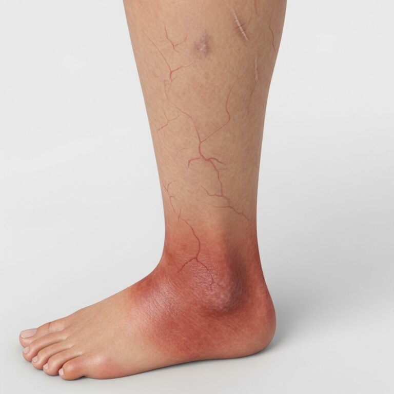
Skin manifestations
Purpura appears as non-blanching macules coalescing into ecchymoses, often on extremities. Purpura fulminans is a severe form with dermal vascular thrombosis leading to necrosis, bullae, and gangrene—requiring urgent debridement. Retiform purpura indicates microvascular occlusion.
Diagnosis of disseminated intravascular coagulation
DIC is a syndrome diagnosed clinically with laboratory confirmation using the International Society on Thrombosis and Haemostasis (ISTH) scoring system. A score ≥5 indicates overt DIC with >90% specificity.
ISTH DIC Score Components
| Parameter | Points |
|---|---|
| Platelet count | >100k: 0; 50-100k: 1; <50k: 2 |
| Elevated fibrin markers (D-dimer) | No increase: 0; Moderate: 2; Strong: 3 |
| Prolonged PT | 3s: 1; 6s: 2; >6s: 3 |
| Fibrinogen | >1g/L: 0; <1g> |
Additional tests: Low factor VIII distinguishes DIC from cirrhosis (where it’s elevated). Dynamic worsening of parameters over hours confirms DIC.
Investigations for disseminated intravascular coagulation
- Full blood count: Thrombocytopenia (platelets <100 x 10^9/L).
- Coagulation profile: Prolonged PT/PTT, low fibrinogen (<1.5 g/L), elevated D-dimer/FDP.
- ISTH score: As above.
- Underlying disease workup: Blood cultures, imaging, tumor markers.
- Skin biopsy (if purpura fulminans): Confirms thrombosis/necrosis.
Treatment of disseminated intravascular coagulation
Primary therapy targets the underlying disorder (e.g., antibiotics for sepsis, chemotherapy for APL). Supportive measures address hemostatic derangements.
| Clinical Scenario | Treatment |
|---|---|
| Acute DIC without bleeding/ischaemia | Observation; treat underlying cause |
| Acute DIC with bleeding | Platelets, FFP, cryoprecipitate (fibrinogen <1.5g/L), PCC |
| Acute DIC with ischaemia | Correct bleeding first, then heparin/LMWH |
| Chronic DIC without thromboembolism | Prophylactic LMWH if high-risk |
| Chronic DIC with thromboembolism | Full anticoagulation (heparin/LMWH) |
Antithrombin concentrates or recombinant activated protein C (if available) for refractory cases. Avoid routine antifibrinolytics due to thrombosis risk.
Complications of disseminated intravascular coagulation
- Microvascular thrombosis: Organ ischemia (kidney, lung, brain).
- Massive hemorrhage: Intracranial, retroperitoneal.
- Multi-organ failure: Mortality 20-50%.
- Limb amputation: In purpura fulminans.
Prevention of disseminated intravascular coagulation
Early recognition of at-risk patients (sepsis, trauma) with prophylactic heparin in select cases. Prompt treatment of triggers reduces incidence.
Timeline and prognosis
Acute DIC evolves over hours-days; mortality correlates with underlying disease severity (e.g., 40% in sepsis). Chronic DIC has better prognosis with anticoagulation. Resolution follows underlying disease control.
Patient follow-up
Monitor coagulation parameters post-treatment. Hepatology referral for liver-related cases. Long-term anticoagulation if chronic form persists.
Frequently Asked Questions
What is the most common cause of DIC?
Sepsis, accounting for up to 50% of cases due to endotoxin-mediated tissue factor expression.
How do you differentiate DIC from cirrhosis coagulopathy?
DIC shows rapid worsening, low factor VIII, high D-dimer; cirrhosis has stable parameters and elevated factor VIII.
Is heparin safe in bleeding DIC patients?
No—correct coagulopathy with blood products first, then use for thrombosis-dominant states.
What is purpura fulminans?
A severe DIC variant with skin necrosis from microvascular thrombosis, often in sepsis or protein C deficiency.
Can DIC resolve completely?
Yes, with treatment of the underlying cause; supportive care restores hemostasis.
References
- Disseminated intravascular coagulation image — DermNet. 2023. https://dermnetnz.org/imagedetail/9662-disseminated-intravascular-coagulation
- Unmasking Disseminated Intravascular Coagulation in Alcohol-Related Cirrhosis — NIH/PMC. 2024-10-15. https://pmc.ncbi.nlm.nih.gov/articles/PMC12558426/
- Disseminated intravascular coagulation — DermNet NZ. 2024. https://dermnetnz.org/topics/disseminated-intravascular-coagulation
- Disseminated Intravascular Coagulation — PubMed Central/NIH. 2019-09-11. https://pmc.ncbi.nlm.nih.gov/articles/PMC6710154/
- Purpura Fulminans — DermNet NZ. 2024. https://dermnetnz.org/topics/purpura-fulminans
Similar Articles


Read full bio of medha deb