Disseminated Secondary Eczema: Guide To Diagnosis & Treatment
Comprehensive guide to understanding, diagnosing, and managing disseminated secondary eczema, a widespread complication of primary skin conditions.

Disseminated secondary eczema is a condition where eczema spreads widely across the body from an initial primary site, often presenting as itchy, inflamed patches due to an idioeccratic response in sensitised individuals.
What is disseminated secondary eczema?
Disseminated secondary eczema, also known as secondary disseminated eczematid or auto-eczematisation, occurs when a primary localised eczema spreads to distant body areas. This happens through an immunological reaction where the initial inflammation triggers a widespread response in previously sensitised skin. It typically arises from chronic primary eczemas like varicose eczema on the lower legs, but can also stem from other dermatoses.
The condition manifests as symmetrically distributed eczematous patches, often intensely pruritic, affecting flexures, trunk, and limbs. Unlike primary eczema, it is not directly contagious but results from hypersensitivity to antigens from the primary site.
Who gets disseminated secondary eczema?
Disseminated secondary eczema affects adults with predisposing primary conditions, particularly those with atopic backgrounds or impaired skin barriers. Common in individuals over 50 with chronic venous insufficiency leading to varicose eczema. Risk factors include:
- Long-standing localised eczema (e.g., varicose, microbial, or discoid).
- History of atopy or contact dermatitis.
- Secondary infections exacerbating the primary site.
Prevalence is higher in patients with poor primary lesion management, where untreated inflammation spreads systemically.
Causes and triggers
The primary cause is a localised eczematous process, most commonly varicose eczema (stasis dermatitis) on the ankles due to venous hypertension. Other triggers include:
- Microbial eczema from bacterial (Staphylococcus aureus) or fungal infections.
- Artefactual eczema or reactions to topical treatments.
- Numerous tiny primary lesions like viral warts or pediculosis.
Pathophysiology involves auto-sensitisation: soluble antigens from damaged primary skin migrate, eliciting a T-cell mediated response elsewhere. Irritants, allergens, or infections amplify dissemination.
Clinical features
Symptoms begin with intense itching at the primary site, followed by dissemination 1-2 weeks later. Key features include:
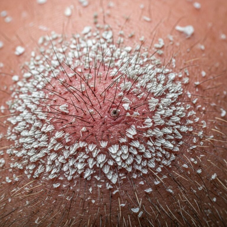
- Symmetrical erythematous patches with vesicles, scaling, and crusting on trunk, limbs, flexures.
- Primary site shows thickened, weepy skin (e.g., gravitational eczema on legs).
- Excoriations from scratching, risk of secondary infection (pustules, oozing).
Skin appears dry, inflamed; severe cases involve erythroderma. Progression: primary inflammation → dissemination → possible autosensitisation nodules.
Diagnosis
Diagnosis is clinical, based on history of primary eczema and dissemination pattern. Essential steps:
- Examine primary site (e.g., lower leg changes in venous eczema).
- Patch testing to exclude contact allergy.
- Swabs/cultures for infection (S. aureus, HSV).
- Biopsy if atypical: spongiosis, acanthosis, parakeratosis.
Differentiate from dermatophytosis, psoriasis, or lymphoma via dermoscopy and histopathology.
Differential diagnosis
| Condition | Key Distinguishing Features |
|---|---|
| Id reaction (to fungi) | Asymmetrical vesicles near primary fungal site; KOH positive. |
| Systemic contact dermatitis | History of allergen exposure; positive patch tests. |
| Drug eruptions | Recent medication start; morbilliform rash. |
| Pre-mycotic eruptions | Follows untreated tinea; annular lesions. |
| Psoriasis | Well-defined plaques, Auspitz sign; nail changes. |
Secondary infections complications
Infection complicates 70-90% of cases due to barrier breach. Common pathogens:
- Bacterial: S. aureus (impetiginisation), streptococci.
- Viral: Eczema herpeticum (HSV).
- Fungal: Candida in flexures.
Signs: crusting, pustules, fever. Treat promptly to prevent dissemination.
Treatment of disseminated secondary eczema
Management targets primary site first, then disseminated areas. Core principles: restore barrier, reduce inflammation, control infection.
General measures
- Emollients: Apply frequently (fragrance-free ointments) to hydrate and protect.
- Avoid irritants: mild soaps, cotton clothing.
- Cool compresses for acute weeping.
Topical treatments
- Corticosteroids: Mid-potency (e.g., mometasone) twice daily for 7-14 days, taper to proactive use.
- Calcineurin inhibitors (tacrolimus) for face/flexures, steroid-sparing.
- Antibiotics: Topical mupirocin for localised infection; oral cephalexin 7-10 days if widespread.
Super-high potency limited to 3 weeks; low-potency no limit.
Treating the primary site
Critical for halting dissemination:
- Varicose eczema: compression therapy, leg elevation.
- Microbial: eradicate infection source (e.g., intranasal mupirocin).
- Venous: address underlying insufficiency.
Systemic therapy
- Antihistamines for itch (non-sedating daytime, sedating night).
- Phototherapy (NB-UVB) for moderate-severe refractory cases.
- Immunosuppressants: Cyclosporine short-term if severe.
Wet wraps with corticosteroid for intensive control.
Complications management
For infected cases: culture-guided antibiotics; avoid TCIs in acute HSV. Monitor for resistance (MRSA).
Prevention
Prevent by aggressive primary site treatment:
- Daily emollients, proactive topicals.
- Compression for venous disease.
- Family staph decolonisation if recurrent.
- Avoid triggers, educate on early intervention.
Outlook
Prognosis excellent with primary control; dissemination resolves in weeks. Chronic if primary persists (e.g., untreated varicose). Relapse risk high without maintenance. Long-term: monitor for venous complications.
Frequently Asked Questions (FAQs)
Can disseminated secondary eczema be cured?
Yes, by treating the primary site effectively; disseminated rash clears once trigger resolved.
Is it contagious?
No, not directly; secondary infections may spread if untreated.
How long does it last?
1-4 weeks with treatment; longer if primary untreated.
What if infection occurs?
Seek antibiotics promptly; culture if no response in 48h.
Best moisturizer?
Fragrance-free ointments like petrolatum-based, applied post-bath.
References
- Treatment of Atopic Dermatitis — American Academy of Pediatrics (AAP). 2023-10-01. https://www.aap.org/en/patient-care/atopic-dermatitis/treatment-of-atopic-dermatitis/
- Atopic dermatitis (eczema) – Diagnosis and treatment — Mayo Clinic. 2024-05-15. https://www.mayoclinic.org/diseases-conditions/atopic-dermatitis-eczema/diagnosis-treatment/drc-20353279
- Eczema – StatPearls — NCBI Bookshelf, NIH. 2024-08-20. https://www.ncbi.nlm.nih.gov/books/NBK538209/
- Eczema management — Royal Children’s Hospital Melbourne. 2024-02-10. https://www.rch.org.au/rchcpg/hospital_clinical_guideline_index/eczema_management/
- Atopic Dermatitis: Diagnosis and Treatment — American Academy of Family Physicians (AAFP). 2020-05-15. https://www.aafp.org/pubs/afp/issues/2020/0515/p590.html
Similar Articles


Read full bio of medha deb