Dyskeratosis Congenita: Causes, Symptoms, and Treatment
Understanding dyskeratosis congenita: A comprehensive guide to this rare genetic bone marrow disorder.

Dyskeratosis Congenita: Understanding a Rare Genetic Disorder
Dyskeratosis congenita (DC) is a rare inherited genetic disorder characterized by defective telomere maintenance and bone marrow failure. This complex condition affects multiple organ systems and presents with a classic triad of abnormal skin pigmentation, nail dystrophy, and oral leukoplakia. The disease manifests differently among patients, ranging from mild to severe presentations, making it a heterogeneous condition that requires individualized management approaches.
What Is Dyskeratosis Congenita?
Dyskeratosis congenita is a genetic form of bone marrow failure, representing the inability of bone marrow to produce sufficient blood cells. The condition is rooted in defective telomere maintenance, leading to premature telomere shortening and subsequent replicative senescence. This results in premature stem cell exhaustion and tissue failure, affecting hematopoietic stem cells and other rapidly dividing cell populations.
The disease can be inherited in three distinct patterns: X-linked, autosomal dominant, or autosomal recessive inheritance. Mutations in at least ten telomere- and telomerase-associated genes have been identified in dyskeratosis congenita, though mutations in known genes only account for approximately one-half of patients with classical clinical manifestations, suggesting additional undiscovered genes are involved.
Clinical Features and Symptoms
Classic Triad
The classic presentation of dyskeratosis congenita includes three primary features:
- Abnormal skin pigmentation: Often presenting as reticulated or lacy patterns on the neck, chest, and thighs
- Nail dystrophy: Characterized by ridging, thinning, and discoloration of fingernails and toenails
- Oral leukoplakia: White patches or plaques in the oral mucosa, increasing cancer risk
These cutaneous and mucocutaneous manifestations may be present before the development of bone marrow failure, sometimes appearing in childhood or early adolescence.
Bone Marrow Failure
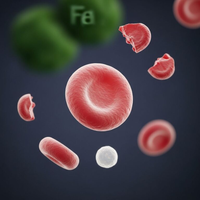
Bone marrow failure is usually diagnosed through observation of low numbers of circulating blood cells, including:
- Red blood cells (leading to anemia)
- White blood cells (resulting in leukopenia)
- Platelets (causing thrombocytopenia)
Individuals with anemia experience tiredness, increased need for sleep, weakness, lightheadedness, dizziness, irritability, headaches, pale skin color, and difficulty breathing. Patients with low white blood cell counts face increased risk of bacterial and fungal infections, while those with low platelet counts are susceptible to excessive bruising and spontaneous bleeding from mucous membranes, particularly the gums and nose.
Additional Systemic Manifestations
Beyond the classic triad, dyskeratosis congenita presents with diverse systemic features affecting multiple organ systems:
- Short stature and growth abnormalities
- Eye abnormalities: Including narrow tear ducts that may become blocked (leading to epiphora), blepharitis, conjunctivitis, ectropion, entropion, and trichiasis
- Dental problems: Cavities, tooth loss, severe periodontal destruction with gingival inflammation and bone loss
- Hair abnormalities: Premature graying and early hair loss
- Pulmonary disease: Interstitial lung fibrosis causing decreased oxygen transport
- Liver disease
- Gastrointestinal abnormalities
- Bone thinning (osteoporosis)
- Infertility and underdeveloped testes (hypogonadism)
- Urinary tract anomalies, particularly hypospadism
- Skeletal abnormalities
- Excessive sweating (hyperhidrosis) of palms and soles
- Esophageal stricture leading to narrowing of the esophagus
- Learning difficulties and developmental delays
An increased incidence of leukemia and cancer, particularly squamous cell carcinoma and hematolymphoid neoplasms, has been well documented in patients with dyskeratosis congenita.
Genetic Basis and Inheritance Patterns
X-Linked Dyskeratosis Congenita
The first gene identified in dyskeratosis congenita was DKC1. Mutations in DKC1 are responsible for the X-linked form and account for approximately 20-25% of sporadic cases. Male patients with DKC1 gene mutations almost always present with the classic form of dyskeratosis congenita, exhibiting high disease penetrance. Mutations in DKC1 have also been found in patients with severe variants of the disease.
De Novo and Sporadic Mutations
In a significant proportion of patients with moderate or severe disease, the mutation is not inherited but has newly arisen either in the germ cells of the parents or shortly after conception. De novo mutations in DKC1 and TINF2 have been found to be responsible for sporadic dyskeratosis congenita, whereas spontaneous mutations in TERT causing disease in the first generation are very rare. Patients with de novo mutations usually have moderate to severe and progressive disease.
Genetic Mechanism
Premature accelerated telomere shortening is thought to be the underlying mechanism of disease pathology. The critical timing at which telomeres become critically short greatly determines the clinical picture. In severe forms of dyskeratosis congenita, telomeres become critically short early in life, in classic forms they become critically short during childhood and adolescence, and in atypical forms they become critically short in adults.
Severe Variants of Dyskeratosis Congenita
Hoyeraal-Hreidarsson Syndrome
Once considered a separate disorder, Hoyeraal-Hreidarsson syndrome (HHS) is now identified as a severe variant of dyskeratosis congenita. Symptoms usually occur during the first year of life and include:
- Severe growth retardation that occurs before birth (intrauterine growth retardation)
- Bone marrow failure
- Immune system deficiencies
- Underdevelopment of the cerebellum (cerebellar hypoplasia)
- Clumsiness caused by inability to coordinate voluntary movements (ataxia)
- Microcephaly (head circumference smaller than expected for infant’s age and sex)
- Moderate to severe mental retardation and developmental delays
A diagnosis of Hoyeraal-Hreidarsson syndrome can be made if the patient has four or more of these features or has cerebellar hypoplasia and additional features of dyskeratosis congenita.
Revesz Syndrome
Similarly, Revesz syndrome, previously known as a distinct disorder, is now understood to share the same underlying abnormality as dyskeratosis congenita and is caused by mutations in the same genes responsible for the classic form. These severe variants represent different clinical manifestations of the same fundamental genetic defect.
Diagnosis of Dyskeratosis Congenita
Diagnosis of dyskeratosis congenita involves a comprehensive clinical and laboratory evaluation. Physicians should look for the classic triad of mucocutaneous features combined with bone marrow failure. Key diagnostic criteria include:
- Complete blood count showing pancytopenia or cytopenia
- Bone marrow examination revealing hypocellularity
- Recognition of characteristic skin findings including reticulated hyperpigmentation
- Identification of nail dystrophy
- Documentation of oral mucosal changes
- Assessment of telomere length using flow cytometry-FISH (fluorescence in situ hybridization)
- Genetic testing for mutations in known dyskeratosis congenita-associated genes
Oral features are particularly important diagnostic indicators, as severe periodontal destruction occurs due to anomalies in ectodermal-derived structures and poor immune response caused by neutropenia. Multiple permanent teeth with decreased root-to-crown ratios may suggest a diagnosis of dyskeratosis congenita.
Treatment and Management Options
Current Therapeutic Approaches
Unfortunately, there is no cure for dyskeratosis congenita currently. Management is dependent on the individual and the progression of their condition. Current therapeutic options are supportive and utilize a multi-disciplinary team approach, including different healthcare professionals to address the unique needs of each individual.
Symptomatic Management
Treatment focuses on managing symptoms and complications, which may include:
- Organ function assessment: Regular monitoring of lung, liver, and kidney function
- Blood count monitoring: Frequent complete blood count assessments
- Androgen therapy: Used in cases of bone marrow failure to stimulate blood cell production
- Supportive blood transfusions: For management of severe anemia and thrombocytopenia
- Lung therapies: Management of pulmonary fibrosis and respiratory complications
- Oral hygiene and periodontal care: Monthly oral self-monitoring for changes in mucous membranes
- Nutritional support and growth monitoring
Important Treatment Considerations
Several important precautions must be observed in the management of dyskeratosis congenita:
- Avoid combining androgens with G-CSF (granulocyte-colony stimulating factor), as this combination is associated with splenic rupture
- Do not collect blood reserves from related donors if a bone marrow transplant is being considered
- Avoid carcinogenic agents including nicotine and alcohol to reduce additional cancer risk
- Treatment protocols should be tailored to the manifestation and severity of disease
Allogeneic Hematopoietic Stem Cell Transplantation
Allogeneic hematopoietic stem cell transplantation (allo-HSCT) is the only curative treatment for bone marrow failure in patients with dyskeratosis congenita. Historically, HSCT was associated with significant morbidity and mortality for these patients. However, the utilization of new transplant regimens with better tolerance is resulting in improved outcomes. Once diagnosed, it is recommended that the patient be managed by a cancer predisposition specialist, particularly for cancer screening and prevention strategies.
Prognosis and Long-Term Complications
The prognosis of dyskeratosis congenita varies significantly based on the severity of disease presentation and the rate of telomere shortening. Patients with dyskeratosis congenita usually die of bone marrow failure due to deficient renewing capability of hematopoietic stem cells. Additionally, the significantly elevated cancer risk, particularly for squamous cell carcinoma and hematologic malignancies, represents a major source of morbidity and mortality in this patient population.
Long-term complications may include progressive pulmonary fibrosis, liver cirrhosis, gastrointestinal complications, and infertility. The multi-system nature of the disease necessitates ongoing surveillance and management of affected organ systems throughout the patient’s lifetime.
Frequently Asked Questions
Q: At what age does dyskeratosis congenita typically appear?
A: The skin changes of dyskeratosis congenita may appear in childhood, while bone marrow failure typically develops during childhood and adolescence. However, severe variants like Hoyeraal-Hreidarsson syndrome may present symptoms within the first year of life. Atypical forms may not manifest until adulthood.
Q: Is dyskeratosis congenita inherited?
A: Dyskeratosis congenita can be inherited in X-linked, autosomal dominant, or autosomal recessive patterns depending on which gene is mutated. However, some cases arise from de novo (new) mutations that occur spontaneously in the germ cells or shortly after conception.
Q: What is the relationship between dyskeratosis congenita and cancer risk?
A: Patients with dyskeratosis congenita have a significantly increased incidence of leukemia and solid cancers, particularly squamous cell carcinoma. This elevated cancer risk is related to the defective telomere maintenance underlying the disease and represents a major source of morbidity and mortality.
Q: Can dyskeratosis congenita be cured?
A: Currently, there is no cure for dyskeratosis congenita. Allogeneic hematopoietic stem cell transplantation is the only curative treatment available specifically for bone marrow failure, though it carries risks. Otherwise, management is supportive and focused on treating complications.
Q: What specialists should manage dyskeratosis congenita?
A: Dyskeratosis congenita requires a multi-disciplinary approach involving hematologists, cancer predisposition specialists, pulmonologists, hepatologists, dermatologists, dentists, and other specialists depending on which organ systems are affected in the individual patient.
References
- Dyskeratosis Congenita – Symptoms, Causes, Treatment — National Organization for Rare Disorders (NORD). 2024. https://rarediseases.org/rare-diseases/dyskeratosis-congenita/
- The diagnosis and treatment of dyskeratosis congenita: a review — García MSF et al. PubMed Central. 2014. https://pmc.ncbi.nlm.nih.gov/articles/PMC4145822/
- Dyskeratosis Congenita — Molecular Cancer Research Centre (MRCC). 2024. https://www.mrcc.org.uk/clinical-diagnostics/other-specialist-clinical-services/chromosome-instability-syndromes/dyskeratosis-congenita/
- Dyskeratosis Congenita Symptoms, Causes & Treatment — Cleveland Clinic. 2024. https://my.clevelandclinic.org/health/diseases/dyskeratosis-congenita
- Dyskeratosis Congenita — Cincinnati Children’s Hospital Medical Center. 2024. https://www.cincinnatichildrens.org/health/d/dyskeratosis-congenita
- Dyskeratosis Congenita in Children — Cedars-Sinai. 2024. https://www.cedars-sinai.org/health-library/diseases-and-conditions—pediatrics/d/dyskeratosis-congenita-in-children.html
- Dyskeratosis congenita — DermNet New Zealand. 2024. https://dermnetnz.org/topics/dyskeratosis-congenita
Similar Articles


Read full bio of medha deb