Ecthyma Gangrenosum Explained: Causes, Diagnosis & Treatment
Critical skin infection linked to Pseudomonas in immunocompromised patients, demanding urgent diagnosis and antibiotics.

What is ecthyma gangrenosum?
Ecthyma gangrenosum is a distinctive and potentially life-threatening cutaneous infection most commonly associated with Pseudomonas aeruginosa bacteraemia in critically ill, immunocompromised patients. It represents a severe dermatological emergency characterised by rapidly progressive necrotic skin lesions that evolve from haemorrhagic pustules into deep ulcers with black eschars. While Pseudomonas aeruginosa remains the primary pathogen, accounting for 60–90% of cases, other opportunistic organisms such as Escherichia coli, Klebsiella species, Staphylococcus aureus, Candida, and various fungi can produce similar ecthyma gangrenosum-like lesions.
The condition arises from bacterial invasion of the vessel walls (vasculitis), leading to thrombosis, infarction, and tissue necrosis with sparse inflammatory response due to the host’s impaired immunity. Ecthyma gangrenosum serves as a critical clinical marker, warranting immediate investigation for systemic sepsis, as mortality rates can exceed 70% in untreated or delayed cases, particularly in neutropenic patients. Early recognition is paramount, as prompt antibiotic therapy can dramatically improve outcomes.
Who gets ecthyma gangrenosum?
Ecthyma gangrenosum predominantly affects individuals with profound immunosuppression, where defects in neutrophil function or number impair bacterial clearance. High-risk groups include:
- Patients with prolonged neutropenia (absolute neutrophil count <500/mm³), often from chemotherapy for haematological malignancies like leukaemia or lymphoma.
- Individuals with advanced HIV/AIDS, particularly CD4 counts <200 cells/µL.
- Transplant recipients on immunosuppressive regimens (solid organ or bone marrow).
- Burn victims with extensive skin barrier loss.
- Neonates, elderly patients, and those with chronic diseases like diabetes mellitus, chronic renal failure, or liver cirrhosis.
- Critically ill patients in intensive care with indwelling catheters, mechanical ventilation, or recent surgery.
Although rare, ecthyma gangrenosum can occur in immunocompetent hosts, typically presenting with localised infection rather than disseminated disease. Risk escalates with prolonged hospital stays, broad-spectrum antibiotic use disrupting normal flora, and invasive procedures.
What causes ecthyma gangrenosum?
Pseudomonas aeruginosa, a ubiquitous Gram-negative, aerobic bacillus thriving in moist environments, is the archetypal causative agent. This opportunistic pathogen exhibits virulence factors including exotoxins, proteases, and biofilm formation that facilitate vascular invasion and tissue destruction. Bacteraemia precedes skin lesions in 37–95% of cases, with haematogenous seeding to dermal vessels.
Alternative pathogens causing ecthyma gangrenosum-like lesions include:
- Gram-negative bacteria: Escherichia coli, Klebsiella pneumoniae, Proteus spp., Serratia marcescens, Burkholderia spp.
- Gram-positive: Staphylococcus aureus (including MRSA), Streptococcus pyogenes
- Fungi: Candida spp., Aspergillus, Mucorales, Fusarium
- Anaerobes and atypical mycobacteria in rare instances
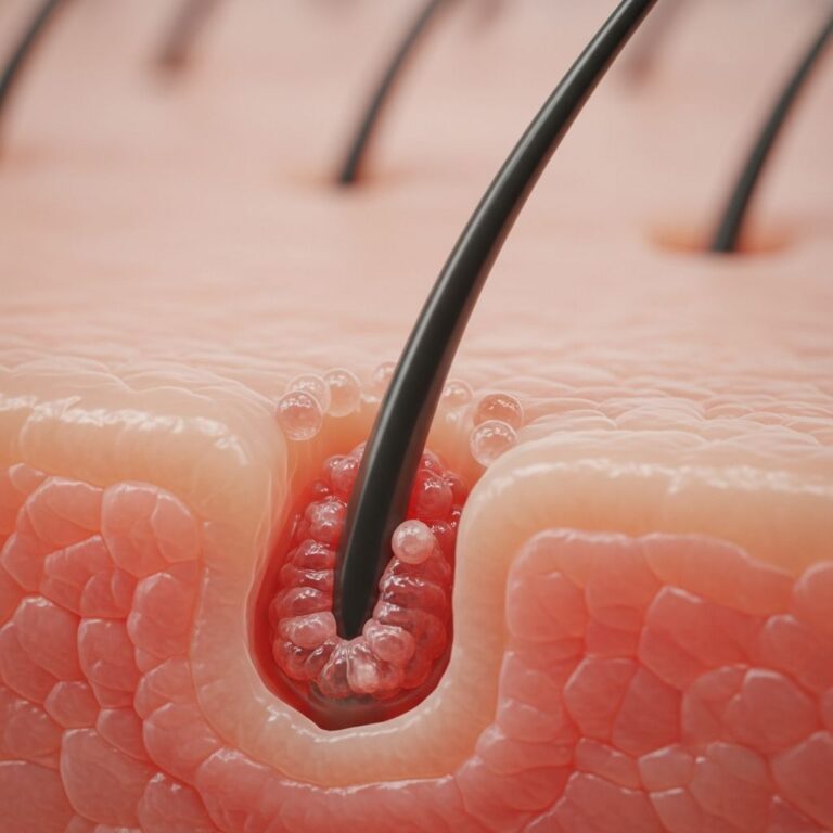
Pathogenesis involves bacterial emboli lodging in dermal vessels, inducing endothelial damage, fibrin thrombi formation, and ischaemic necrosis with minimal leukocytic infiltration.
What are the clinical features of ecthyma gangrenosum?
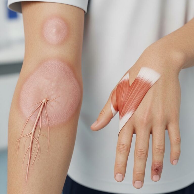
Lesions typically emerge in flexural or perineal areas (axillae, groin, buttocks) but can appear anywhere, including trunk and extremities. The evolution is remarkably rapid:
- Initial phase (0–24 hours): Painless erythematous macules or patches (1–15 mm), indurated with surrounding oedema.
- Pustular phase: Central vesicle or haemorrhagic pustule forms, becoming bullous with purple discolouration.
- Necrotic phase (12–24 hours): Blister ruptures, revealing a grey-black eschar (2–20 cm) with erythematous halo; punched-out ulcer with undermined edges.
Multiple lesions (average 3–10) may develop sequentially over days, often with systemic signs: fever, chills, hypotension, altered mental status indicating sepsis. Lesions are initially non-tender but become painful as necrosis advances. In non-bacteraemic cases, solitary lesions predominate without systemic features.
How is ecthyma gangrenosum diagnosed?
Diagnosis combines clinical suspicion, microbiology, and histopathology.
Clinical assessment
Suspect ecthyma gangrenosum in immunocompromised patients with rapidly evolving necrotic lesions, especially during neutropenic fever.
Microbiological tests
- Blood cultures: Positive in 37–95%; take 2–3 sets during fever spikes.
- Wound swabs/biopsy: From vesicle fluid, ulcer base, or under eschar; Gram stain shows Gram-negative rods.
- Tissue culture: For bacteria, fungi, mycobacteria; sensitivity testing essential.
Histopathology
Skin biopsy reveals pauci-inflammatory vascular necrosis with intravascular Gram-negative bacilli, thrombosis, haemorrhage, and dermal infarction. Special stains (Gram, GMS, PAS, AFB) identify organisms.
Additional tests
- Complete blood count, CRP, procalcitonin, lactate for sepsis assessment.
- Imaging (CT/MRI) if deep extension suspected.
- Wood’s lamp: Pseudomonas fluoresces green.
What is the differential diagnosis for ecthyma gangrenosum?
| Condition | Key Distinguishing Features |
|---|---|
| Pyoderma gangrenosum | Preceded by pathergy; more inflammatory; biopsy shows neutrophilic infiltrate without organisms |
| Calciphylaxis | Renal failure patients; livedo pattern; calcium oxalate deposits on biopsy |
| Aspergillosis/mucormycosis | Branching hyphae on biopsy; sinus involvement |
| Septic emboli (endocarditis) | Multiple peripheral lesions; heart murmur; positive blood cultures |
| Antibacterial drug reactions (e.g., vancomycin) | History of recent antibiotics; resolves with discontinuation |
| Cutis marmorata telangiectatica congenita | Congenital, persistent reticulate erythema; no necrosis |
What is the treatment for ecthyma gangrenosum?
Initiate empirical broad-spectrum IV antibiotics immediately after cultures, targeting Pseudomonas.
Antimicrobial therapy
- First-line: Piperacillin-tazobactam 4.5g IV Q6H or cefepime 2g IV Q8H ± vancomycin for MRSA coverage.
- Alternatives: Ceftazidime, imipenem, meropenem, or ciprofloxacin + piperacillin (double Pseudomonas coverage).
- De-escalate based on sensitivities; duration 2–6 weeks.
Surgical intervention
Wide debridement of necrotic tissue ± abscess drainage; consider skin grafting for large defects. Healing by secondary intention or VAC therapy viable.
Supportive care
- Reverse neutropenia (G-CSF if <500 neutrophils).
- ICU management for septic shock.
- Wound care: Silver sulfadiazine, mupirocin.
What is the outcome for ecthyma gangrenosum?
Mortality 10–70%, highest in bacteraemic/neutropenic patients despite treatment. Survivors may have scarring/atrophic ulcers. Early antibiotics improve survival to >80%. Relapse occurs in 10–20% with persistent immunosuppression.
Frequently asked questions about ecthyma gangrenosum
Q: Is ecthyma gangrenosum contagious?
A: No. It results from systemic/opportunistic infection in immunocompromised hosts, not person-to-person transmission.
Q: Can ecthyma gangrenosum occur in healthy people?
A: Rarely, as localised infection following trauma or folliculitis; lacks bacteraemia.
Q: How quickly do ecthyma gangrenosum lesions progress?
A: Macule to necrotic ulcer in 12–24 hours; emergency requiring immediate intervention.
Q: What antibiotics treat Pseudomonas ecthyma gangrenosum?
A: Anti-pseudomonal beta-lactams (pip-tazo, cefepime) ± aminoglycoside; tailor to sensitivities.
Q: Does surgery always require for ecthyma gangrenosum?
A: Indicated for extensive necrosis/abscesses; medical therapy suffices for small, superficial lesions.
References
- Ecthyma gangrenosum – DermNet — DermNet NZ. 2024-07. https://dermnetnz.org/topics/ecthyma-gangrenosum
- Diagnosis and Management of Ecthyma Gangrenosum in Chronic Renal Failure — PMC / Eplasty. 2014-05-28. https://pmc.ncbi.nlm.nih.gov/articles/PMC4037781/
- Ecthyma Gangrenosum – MD Searchlight — MD Searchlight. 2025. https://mdsearchlight.com/infectious-disease/ecthyma-gangrenosum/
- Ecthyma Gangrenosum – StatPearls — NCBI Bookshelf. 2023-07-17. https://www.ncbi.nlm.nih.gov/books/NBK534777/
- Ecthyma (including ecthyma gangrenosum) — PCDS. 2024. https://www.pcds.org.uk/clinical-guidance/ecthyma-gangrenosum
Similar Articles


Read full bio of Sneha Tete