Elastosis Perforans Serpiginosa: 4 Treatment Options & Symptoms
Rare skin disorder featuring transepidermal elimination of abnormal elastic fibres in serpiginous patterns on neck and limbs.

Elastosis perforans serpiginosa (EPS) is a rare perforating dermatosis characterised by transepidermal elimination of abnormal elastic fibres from the papillary dermis through the epidermis, presenting as grouped erythematous papules in annular or serpiginous patterns.
Introduction
Elastosis perforans serpiginosa belongs to the group of perforating dermatoses, where altered dermal components are extruded through the epidermis. This process, known as transepithelial or transepidermal elimination, occurs when the epidermis perceives the abnormal elastic tissue as foreign material, triggering an inflammatory response that facilitates its removal. EPS is distinct from other elastotic conditions like solar elastosis, which involves diffuse dermal degeneration without perforation.
The condition was first described in the mid-20th century and remains uncommon, with fewer than 300 cases reported in medical literature. It manifests clinically as small, hyperkeratotic papules arranged in characteristic snake-like (serpiginous) or arciform configurations, primarily on the neck and upper limbs. While generally asymptomatic, mild pruritus can occur, and spontaneous resolution is common within months to years.
Demographics
EPS most frequently emerges in early adulthood, with a peak incidence between 20 and 30 years of age, though cases have been documented in children as young as 5 years and in elderly patients over 70. There is a marked male predominance, with a male-to-female ratio of approximately 4:1, suggesting possible hormonal or genetic influences.
Racial distribution appears universal, with no strong predilection for specific ethnic groups, though underreporting in certain populations may skew data. Familial cases are rare but reported, particularly in association with inherited connective tissue disorders.
Causes
The pathogenesis of EPS involves focal degeneration and perforation of abnormal elastic fibres in the papillary dermis. Three main forms are recognised:
- Idiopathic EPS: Occurs without underlying systemic disease or external triggers. Represents about 20-25% of cases and may stem from sporadic genetic mutations affecting elastin synthesis or structure.
- Reactive EPS: Associated with inherited connective tissue disorders, including Ehlers-Danlos syndrome (types IV and VII), Marfan syndrome, osteogenesis imperfecta, and pseudoxanthoma elasticum. In these, pre-existing elastic fibre abnormalities predispose to perforating reactions.
- Drug-induced EPS: Most commonly linked to long-term penicillamine therapy, used in Wilson’s disease and cystinuria. Penicillamine alters elastic fibre cross-linking, leading to perpendicular orientation and transepidermal elimination. Onset typically occurs after 1-3 years of treatment, affecting up to 1% of patients on the drug.
Other implicated factors include D-penicillamine alternatives like bucillamine and environmental insults, though these are less common. Histologically, the elastic fibres are thickened, clumped, and brightly eosinophilic, prompting epidermal hyperplasia and channel formation for elimination.
Signs and Symptoms
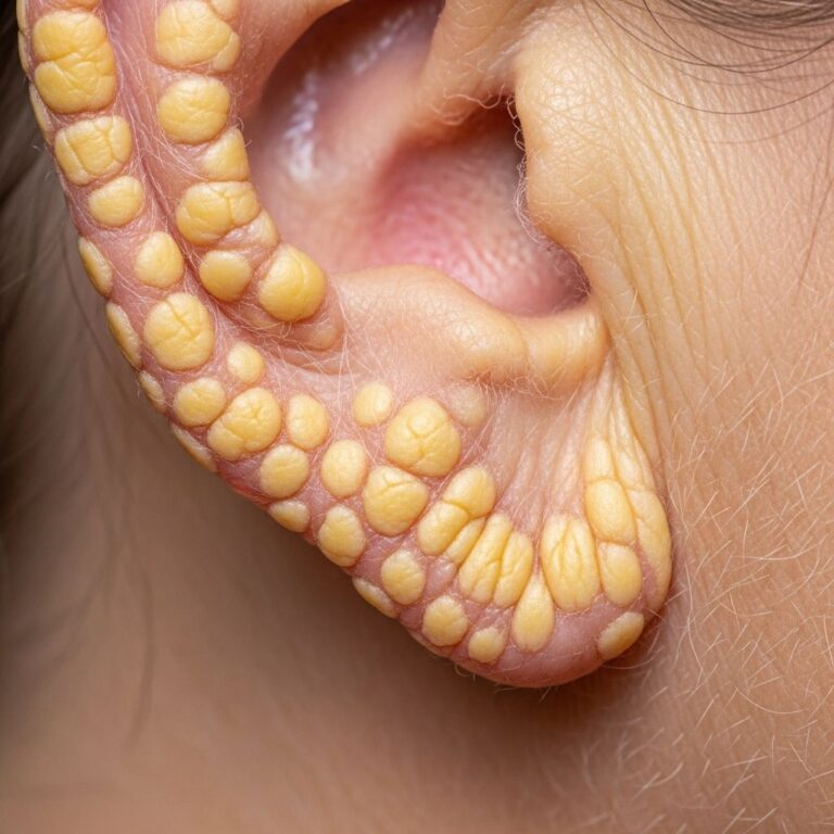
Clinically, EPS presents as clusters of 2-5 mm erythematous or hyperpigmented papules, often with central umbilication, keratotic plugs, or crusting. Lesions arrange in linear, annular, arcuate, or serpiginous patterns, mimicking a snake’s trail—hence the name “serpiginosa.” New papules migrate outward while central ones heal with atrophic scarring, creating expanding configurations up to 7-10 cm in diameter.
Common sites include:
- Neck (70% of cases, often posterior cervical region)
- Upper extremities (20%, forearms and arms)
- Face (11%, perioral or cheek areas)
- Lower limbs (6%, thighs or legs)
- Trunk (3%, less frequent)
Symptoms are usually absent, but koebnerisation (induction by trauma) can occur, and mild to moderate itchiness affects 20-30% of patients. Rarely, lesions cause cosmetic concern or secondary infection.
Diagnosis
Diagnosis relies on characteristic clinical morphology and confirmed by skin biopsy. Dermoscopy may reveal central keratin plugs with surrounding telangiectasia, aiding in-office assessment.
Histopathology: Key features include epidermal invagination filled with basophilic debris, neutrophilic infiltrate, and parakeratosis. Vertically oriented, brightly eosinophilic elastic fibres traverse the epidermis, confirmed by Elastic-Van Gieson (EVG) stain showing black fibres in channels. Dermal changes feature elastic fibre reduplication and loss.
Differential diagnoses include:
| Condition | Key Distinguishing Features |
|---|---|
| Reactive Perforating Collagenosis | Collagen fibres extruded (EVG negative); associated with diabetes/renal failure |
| Kyrle Disease | Hyperkeratotic plugs with inflammatory debris (no elastic fibres) |
| Perforating Folliculitis | Follicular-based; mixed collagen/elastin |
| Traumatic Perforation | Collagen fibres post-trauma (EVG negative); epidermal erosion clues |
Systemic evaluation for connective tissue disorders (e.g., echocardiography for Marfan) or penicillamine use is essential.
Treatment
No curative therapy exists; management targets symptom relief and lesion clearance, as spontaneous remission occurs in 6 months to 5 years for most cases (80-90%).
- Topical therapies: Corticosteroids (e.g., clobetasol), retinoids (tretinoin), and keratolytics (urea, salicylic acid) to reduce hyperkeratosis and inflammation. Success in 40-60% of mild cases.
- Physical modalities: Cryotherapy, curettage, electrodessication, or laser ablation (CO2/Erbium:YAG) for symptomatic or persistent lesions. Effective but risks scarring.
- Systemic options: Allopurinol (300-600 mg/day) induces lesion regression in idiopathic/reactive forms via uric acid modulation of elastolysis; response in 2-4 months. Discontinue penicillamine if drug-induced, with resolution post-cessation.
- Other: Intralesional steroids, imiquimod, or photodynamic therapy as adjuncts.
Monitoring for underlying conditions is crucial; multidisciplinary care with rheumatology/genetics for reactive forms.
Frequently Asked Questions
Is elastosis perforans serpiginosa contagious?
No, EPS is not infectious or contagious; it results from intrinsic elastic tissue abnormalities.
Does EPS always require biopsy for diagnosis?
Classic serpiginous papules on the neck suggest EPS, but biopsy confirms by demonstrating elastic fibre elimination.
Can EPS be prevented?
Idiopathic cases cannot; drug-induced forms may be mitigated by monitoring penicillamine patients and early discontinuation.
How long does elastosis perforans serpiginosa last?
Most resolve spontaneously within 1-3 years, though some persist longer or recur.
Is elastosis perforans serpiginosa linked to skin cancer?
No direct association; it is benign, but chronic lesions warrant monitoring for secondary changes.
References
- Elastosis perforans serpiginosa pathology image — DermNet NZ. 2010. https://dermnetnz.org/imagedetail/12251-elastosis-perforans-serpiginosa-pathology
- Elastosis perforans serpiginosa pathology — DermNet NZ, Dr Ben Tallon. 2010. https://dermnetnz.org/topics/elastosis-perforans-serpiginosa-pathology
- Elastosis perforans serpiginosa — DermNet NZ, Vanessa Ngan. 2003 (updated). https://dermnetnz.org/topics/elastosis-perforans-serpiginosa
- Elastosis — DermNet NZ, Dr Beth Wright. 2012. https://dermnetnz.org/topics/elastosis
Similar Articles


Read full bio of Sneha Tete