Endoscopic Submucosal Dissection: Advanced GI Tumor Removal
Learn about ESD, a minimally invasive procedure for removing gastrointestinal tumors and polyps.

Understanding Endoscopic Submucosal Dissection
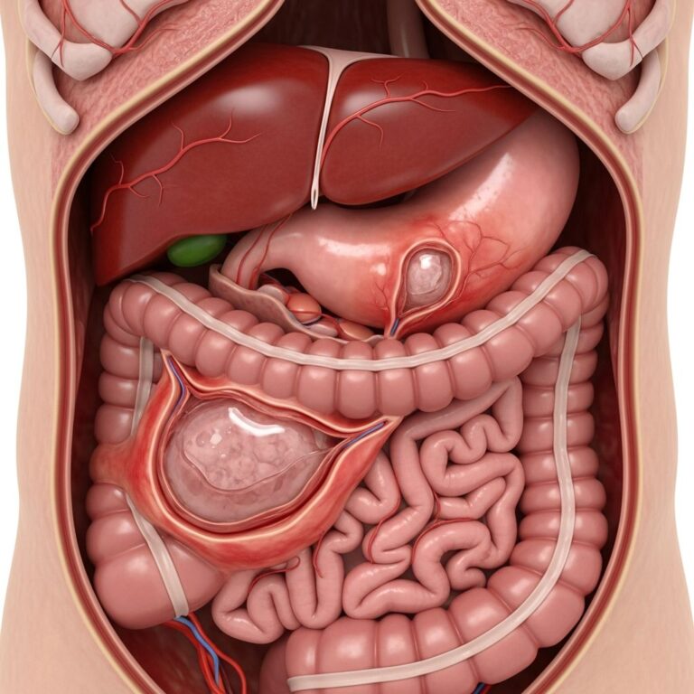
Endoscopic submucosal dissection (ESD) is an advanced minimally invasive procedure designed to remove large polyps, benign growths, and early-stage cancerous lesions from the gastrointestinal tract. Unlike traditional open surgery, ESD allows physicians to treat gastrointestinal neoplasms while preserving the natural anatomy and function of the digestive system. This technique has been extensively used in Asia for many years and is rapidly gaining acceptance in Western medical centers due to its effectiveness and patient-friendly recovery profile.
The procedure is particularly valuable for treating lesions that are confined to the superficial layers of the gastrointestinal tract, specifically those that have not invaded the muscle layer beneath the mucosa. ESD can be performed in multiple locations within the digestive system, including the esophagus, stomach, small intestine, and colon. By enabling complete removal of larger lesions in a single piece—known as en bloc resection—ESD offers significant advantages over conventional polypectomy or endoscopic mucosal resection (EMR) for managing complex gastrointestinal pathology.
When ESD is Recommended
Endoscopic submucosal dissection may be recommended in several clinical scenarios. The procedure is typically indicated for removing flat growths, large polyps exceeding 20 millimeters in diameter, and early-stage cancers that remain confined to the mucosal layer of the gastrointestinal tract. ESD is particularly beneficial when conventional snare-based techniques cannot optimally and radically remove lesions, especially in the colon and rectum where high suspicion of limited submucosal invasion exists.
The most common lesions treated with ESD include tumors of the stomach, esophagus, and colon. For patients with large growths in the rectum, ESD may help avoid permanent colostomy bags and other life-altering surgical consequences. Additionally, ESD can be applied to complex neoplasms such as ulcerative non-lifting neoplasms, recurrent neoplasms, and lesions with submucosal fibrosis that might otherwise be considered surgically challenging or untreatable through less invasive means.
The ESD Procedure: Step-by-Step Process
Understanding how endoscopic submucosal dissection is performed helps patients prepare mentally and physically for the procedure. The technique involves a carefully orchestrated sequence of steps designed to safely and completely remove the lesion while minimizing complications and preserving healthy tissue.
Preparation and Access
The procedure begins with patient preparation and anesthesia. For lesions in the esophagus, stomach, and upper intestine, the endoscope is inserted through the mouth and advanced down the throat. For colonic or rectal lesions, the endoscope is inserted through the anus. The endoscope is a sophisticated instrument consisting of a narrow, flexible tube equipped with a high-definition camera, light source, and working channels through which specialized surgical instruments can be passed.
Marking the Lesion
The first step of the actual procedure involves marking the margins of the lesion that requires removal. The tip of the specialized knife is used to create demarcation lines around the entire perimeter of the abnormal tissue. This marking is critical as it establishes the boundaries for the subsequent dissection and ensures that an adequate margin of healthy tissue is removed along with the lesion to reduce the risk of residual disease.
Submucosal Injection
Once the lesion margins are clearly marked, a solution is injected into the submucosa—the layer of tissue immediately beneath the mucosa. This injection lifts and separates the mucosal layer containing the lesion from the underlying muscular layer. Various injection solutions may be used, each with specific advantages. Sodium chloride (0.9%) is economical and widely available but is rapidly absorbed. Hypertonic saline (3%-4.7%), dextrose solutions (20%-50%), and hydroxypropyl methylcellulose provide more prolonged submucosal elevation, though they may cause local inflammation depending on their osmolality.
Circumferential Incision
After adequate submucosal lifting is achieved, an incision is made around the complete circumference of the lesion using specialized electrosurgical knives. Multiple knife options are available to specialists performing ESD, including the insulation-tipped diathermic knife (IT-knife), needle knife, hook knife, flex knife, triangle-tipped knife, and flush knife. The choice of knife depends on the specific characteristics of the lesion and the anatomical location.
Submucosal Dissection
The final and most technically demanding step involves carefully dissecting the connective tissue within the submucosa to separate the entire lesion from the muscular layer beneath. This step requires meticulous technique to avoid perforation of the muscle layer, which can lead to serious complications. The gastroenterologist uses careful knife movements and sometimes employs traction techniques to improve visualization of the dissection plane.
Advantages of ESD Over Conventional Methods
Endoscopic submucosal dissection offers several significant advantages compared to traditional polypectomy and endoscopic mucosal resection. Understanding these benefits helps explain why this technique is increasingly favored for appropriate lesions.
Complete En Bloc Resection
One of the primary advantages of ESD is the ability to achieve en bloc resection—removing the entire lesion as a single intact specimen—even for large neoplasms. In contrast, conventional EMR often requires piecemeal removal of larger lesions, which significantly increases the risk of local cancer recurrence and makes adequate histopathological staging impossible.
Superior Histopathological Assessment
Because ESD removes the lesion intact with surrounding healthy margin, pathologists can thoroughly examine the specimen to determine whether all abnormal tissue has been removed and whether deeper invasion exists. This complete histopathological assessment is crucial for appropriate staging and predicting prognosis, information that cannot be reliably obtained from fragmented specimens.
Lower Recurrence Rates
The en bloc resection capability and more complete removal of lesions result in significantly lower local recurrence rates compared to EMR or polypectomy. This means patients are less likely to require repeat procedures or develop recurrent disease in the same location.
Resection of Complex Lesions
ESD can successfully treat lesions that would otherwise be considered unresectable or extremely difficult to manage with conventional endoscopic techniques. These include large neoplasms, ulcerative non-lifting neoplasms, recurrent lesions following failed prior resections, and lesions with submucosal fibrosis.
Specialized Tools and Equipment
The success and safety of ESD depend significantly on the availability and proper use of specialized equipment designed to facilitate the procedure. Modern ESD utilizes numerous cutting instruments, visualization aids, injection solutions, and hemostatic devices.
Cutting and Dissection Instruments
A wide array of electrosurgical knives has been developed for ESD, each with specific design characteristics suited to particular situations. These include the IT-knife, needle knife, hook knife, flex knife, triangle-tipped knife, and flush knife. Additionally, specialized devices such as small-caliber tip transparent (ST) hoods are employed to improve visualization during dissection.
Traction Devices
A unique challenge in endoscopic surgery is the lack of a second hand to provide counter-traction during dissection. Various traction methods have been developed to address this limitation, with studies demonstrating that these techniques result in faster procedures, higher resection rates, and lower complication rates. The Endolifter, for example, is a specialized cap with grasping forceps at its distal end that facilitates mucosal elevation and easier dissection while reducing procedure time and bleeding.
Electrosurgical Generators and Coagulation Modes
Advanced electrosurgical generators provide multiple cutting and coagulation modes to optimize tissue management during ESD. These include Swift Coag (similar to Dry Cut with enhanced hemostasis), Forced Coag (higher voltage for deeper dissection), Spray Coag (non-contact surface coagulation for post-procedure hemostasis), and Soft Coag (low voltage coagulation without cutting). Modern generators also offer bipolar capabilities and specialized modes like RFCoag with acoustic resistance feedback for controlled deep tissue coagulation.
The ESD Procedure Timeline and Recovery
Understanding what to expect during and after ESD helps patients prepare appropriately and manage recovery expectations.
Procedure Duration
Endoscopic submucosal dissection typically requires between one to three hours to complete, depending on lesion size, location, complexity, and individual anatomical factors. This is considerably longer than standard polypectomy but significantly shorter than traditional surgical approaches requiring general anesthesia and hospital stays.
Specimen Analysis
Following successful removal, the resected tissue specimen is sent to the laboratory for pathological analysis. A pathologist examines the tissue sample to identify any signs of disease, assess margins, determine invasion depth, and provide staging information critical for determining whether additional treatment is necessary.
Recovery and Hospital Stay
After completion of ESD, patients are transferred to a recovery room where they remain until the effects of anesthesia wear off. Many patients can return home the same day, though some may require an overnight hospital stay depending on the specific procedure performed and individual patient factors. Patients should arrange for someone to provide transportation, as it may take up to 24 hours for the complete effects of anesthesia to resolve fully.
Comparing ESD with Endoscopic Mucosal Resection
While both ESD and EMR are minimally invasive endoscopic procedures, they have important differences that affect their appropriate clinical application.
| Feature | ESD | EMR |
|---|---|---|
| Lesion Size Capability | Effective for lesions >20mm; can remove very large lesions | Limited to smaller lesions; requires piecemeal resection for large lesions |
| Resection Type | En bloc resection (complete removal in single piece) | Often piecemeal removal |
| Recurrence Risk | Lower recurrence rates | Higher recurrence rates with large lesions |
| Histopathology | Complete assessment of margins and invasion depth | Limited assessment, particularly with fragmented specimens |
| Procedure Time | Longer (1-3 hours) | Shorter |
| Complexity | More technically challenging | More straightforward technique |
| Complications | Higher rate of complications | Lower complication rates |
Potential Benefits for Patients
Endoscopic submucosal dissection offers patients numerous advantages that can significantly improve their quality of life and health outcomes.
Avoidance of Major Surgery
Perhaps the most significant benefit is that ESD can help patients avoid major surgical procedures such as removal of the esophagus, stomach, or significant portions of the colon. This translates directly into faster recovery, reduced operative risk, and preservation of normal gastrointestinal anatomy and function.
Elimination of Permanent Ostomies
For patients with large rectal growths, ESD may allow complete lesion removal while avoiding the need for a permanent colostomy bag. The ability to preserve normal bowel continuity and function is profoundly important to patients’ physical and psychological well-being.
Faster Recovery
Compared to open surgery, ESD offers a quicker and less painful recovery period. Many patients can return home the same day and resume normal activities within days rather than weeks or months.
Preserved Gastrointestinal Function
By maintaining the natural anatomy and structure of the gastrointestinal tract, ESD preserves normal digestive function and avoids the long-term nutritional and digestive complications sometimes associated with surgical resection.
Special Expertise and Referral Considerations
Endoscopic submucosal dissection is a technically sophisticated procedure requiring substantial expertise and specialized training. Patients seeking ESD should ensure they are evaluated and treated by gastroenterologists or interventional endoscopists with specific training and experience in ESD techniques. Many academic medical centers and specialized gastrointestinal cancer programs have developed particular expertise in this field.
Frequently Asked Questions
Q: Is ESD covered by insurance?
A: Most insurance plans cover ESD when it is medically necessary for treating appropriate lesions. Patients should verify coverage with their insurance company and healthcare provider before the procedure.
Q: What are the main risks of ESD?
A: While ESD is minimally invasive, potential complications can include perforation of the gastrointestinal wall, post-procedure bleeding, and infection. However, serious complications are relatively uncommon when performed by experienced specialists. Discuss specific risks with your healthcare provider.
Q: Will I need additional treatment after ESD?
A: Additional treatment depends on the pathology results. If cancer is found with concerning features, additional therapy such as radiation or chemotherapy may be recommended. Your medical team will discuss these options based on your specific situation.
Q: How long does recovery take after ESD?
A: Most patients recover significantly within a few days. Complete resolution of anesthesia effects typically occurs within 24 hours. Return to normal activities is usually possible within one to two weeks.
Q: Can ESD be used for all gastrointestinal lesions?
A: No, ESD is specifically indicated for lesions confined to the mucosal and superficial submucosal layers without invasion into the muscle layer or evidence of nodal metastasis. Your gastroenterologist will determine if you are a suitable candidate.
References
- Endoscopic submucosal dissection — Wikipedia. Retrieved from https://en.wikipedia.org/wiki/Endoscopic_submucosal_dissection
- Endoscopic submucosal dissection: an update on tools and techniques — National Center for Biotechnology Information (NCBI). 2020. Retrieved from https://pmc.ncbi.nlm.nih.gov/articles/PMC7545765/
- Endoscopic Submucosal Dissection (ESD) Treatment for GI Cancer — Mercy Medical Center. Retrieved from https://mdmercy.com/mercy-services/treatments/endoscopic-submucosal-dissection
- Endoscopic Submucosal Dissection (ESD): Procedure & Recovery — Cleveland Clinic. Retrieved from https://my.clevelandclinic.org/health/treatments/21091-endoscopic-submucosal-dissection-esd-for-esophageal-and-gastric-cancer
Similar Articles


Read full bio of Sneha Tete