Eosinophilic Fasciitis: Symptoms, Causes, Treatment Guide
Rare inflammatory disorder causing skin thickening, pain, and fasciitis—learn about symptoms, diagnosis, and effective treatments.

Authoritative facts about eosinophilic fasciitis: what it is, symptoms, causes, diagnosis, treatment, and prognosis.
What is eosinophilic fasciitis?
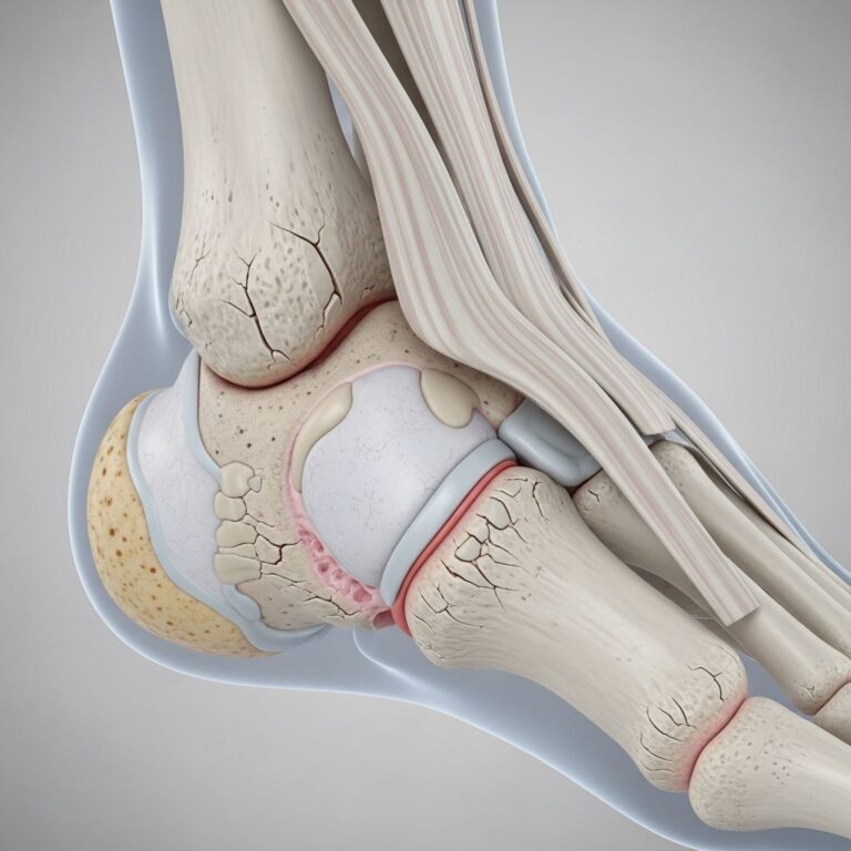
Eosinophilic fasciitis (EF), also known as Shulman syndrome, is a rare fibrosing disorder characterised by oedema and subsequent induration of the skin and subcutaneous tissues, primarily affecting the fascia—the tough connective tissue enveloping muscles, vessels, and nerves. This inflammation leads to painful swelling, skin hardening, and potential joint restriction, most commonly in the arms and legs of middle-aged adults.
The fascia thickens due to infiltration by eosinophils (a type of white blood cell) and subsequent fibrosis, distinguishing EF from scleroderma despite similar skin changes. Unlike scleroderma, EF spares the fingers and toes, and Raynaud phenomenon is absent. The condition often presents symmetrically, with acute onset following triggers like strenuous exercise.
Who gets eosinophilic fasciitis?
Eosinophilic fasciitis predominantly affects adults aged 30–60 years, with a slight male predominance (male-to-female ratio approximately 1.5:1). It is extremely rare, with incidence estimated at less than 1 in 100,000 people annually. Children and elderly individuals are rarely affected.
- Demographics: Middle-aged adults; more common in males.
- Risk factors: History of intense physical exertion (reported in up to 50% of cases), certain medications, or haematologic disorders.
What causes eosinophilic fasciitis?
The exact aetiology of eosinophilic fasciitis remains unknown, but it is considered an autoimmune disorder where eosinophils release cytokines like transforming growth factor-beta (TGF-β), promoting fibrosis in the fascia. Possible triggers include:
- Unusually intense physical activity or trauma (50% of cases).
- Infections (e.g., Borrelia burgdorferi).
- Medications (e.g., atorvastatin, simvastatin, phenytoin, immune checkpoint inhibitors).
- Radiation therapy or graft-versus-host disease.
- Associated haematologic malignancies (e.g., leukaemia, myeloproliferative disorders).
- Environmental exposures or toxins (speculative).
No single genetic predisposition has been identified, though immune dysregulation plays a central role.
What are the clinical features of eosinophilic fasciitis?
Symptoms develop acutely over days to weeks, starting with localised oedema and pain in the extremities. Key clinical features include:
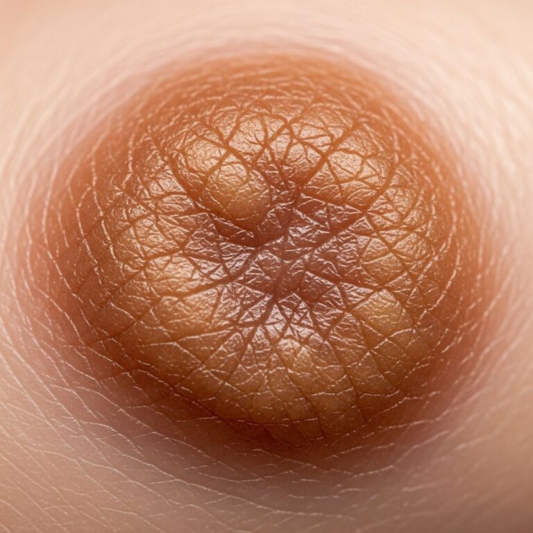
- Skin changes: Symmetrical swelling, induration (peau d’orange or orange-peel appearance), thickened ‘groove sign’ (indentations along superficial veins), and shiny, taut skin. Spares hands, feet, and face.
- Pain and stiffness: Deep muscle pain, tenderness, and restricted joint mobility due to fascial thickening.
- Systemic symptoms: Fatigue, weight loss, fever (infrequent).
- Late features: Joint contractures, muscle weakness, carpal tunnel syndrome.
Physical examination reveals non-pitting oedema and fibrosis, with limited range of motion in affected limbs.
How is eosinophilic fasciitis diagnosed?
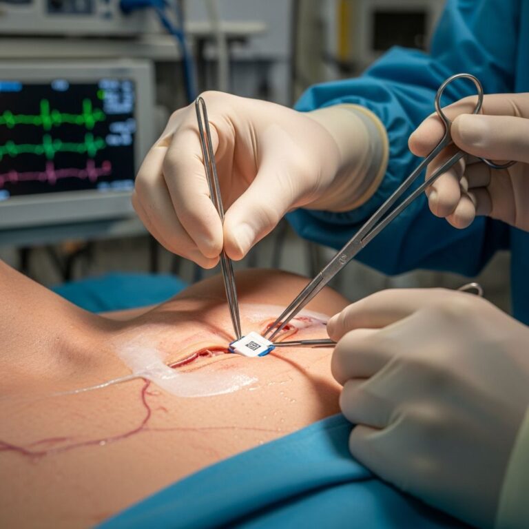
Diagnosis combines clinical findings, laboratory tests, imaging, and histopathology. There is no single confirmatory test, but deep incisional biopsy is gold standard.
Clinical suspicion
Based on characteristic swelling, induration, and history of exertion.
Laboratory tests
- Peripheral
eosinophilia
(50–90% of cases, often marked early). - Elevated erythrocyte sedimentation rate (ESR) and C-reactive protein (CRP).
- Hypergammaglobulinaemia; normal creatine kinase (CK) distinguishes from myositis.
Imaging
- MRI: Thickened fascia (>2 mm signal intensity on T2-weighted images), ‘fascia sign’. Highly sensitive.
- Ultrasound: Fascial thickening and oedema.
Histopathology
Deep skin biopsy (including fascia) shows:
- Inflammatory infiltrate with eosinophils, lymphocytes, plasma cells.
- Fibrosis and collagen deposition in fascia (spares dermis/epidermis).
Differential diagnosis includes scleroderma, eosinophilic myalgia syndrome, and hypereosinophilic syndrome.
Complications of eosinophilic fasciitis
Untreated EF can lead to permanent morbidity:
- Joint contractures and ankylosis limiting mobility.
- Non-erosive arthritis.
- Carpal tunnel syndrome (median nerve compression).
- Compartment syndrome (rare, due to fascial pressure).
- Associated aplastic anaemia or malignancies (5–10% of cases).
What is the treatment for eosinophilic fasciitis?
Treatment should commence promptly post-diagnosis to prevent fibrosis. First-line is high-dose systemic corticosteroids; 80–90% respond.
First-line therapy
- Prednisone: 1 mg/kg/day orally, tapered over 6–24 months based on response.
- Pulse methylprednisolone: 1 g IV for 3 days in severe cases, yielding rapid improvement.
Second-line agents (steroid-sparing)
Add if incomplete response or relapse:
- Methotrexate (15 mg/week).
- Other DMARDs: hydroxychloroquine, ciclosporin, mycophenolate mofetil.
- Immunosuppressants: azathioprine, cyclophosphamide (refractory cases).
- Biologics: rituximab, infliximab (limited evidence).
Supportive measures
- NSAIDs for pain/swelling.
- Physical therapy to maintain joint mobility.
- Moisturisers for skin care.
| Treatment Tier | Agents | Dose/Duration | Response Rate |
|---|---|---|---|
| First-line | Prednisone | 1 mg/kg/day, taper 6–24 months | 80–90% |
| Pulse IV | Methylprednisolone | 1 g/day x 3 days | Rapid in severe cases |
| Second-line | Methotrexate | 15 mg/week | Steroid-sparing |
Prognosis for eosinophilic fasciitis
With early treatment, most achieve remission within 1–3 years; complete resolution in 80–90%. Spontaneous remission occurs in 10–20% after 2–5 years. Relapses possible during steroid taper (20–30%). Poor prognostic factors: delayed diagnosis, associated malignancy.
Long-term sequelae (e.g., contractures) minimised by prompt therapy. Regular monitoring of eosinophil count, inflammatory markers, and MRI guides management.
Frequently Asked Questions (FAQs)
Q: Is eosinophilic fasciitis the same as scleroderma?
A: No. EF affects fascia only, spares fingers/toes, lacks Raynaud’s; scleroderma involves dermis/vessels with systemic features.
Q: Can eosinophilic fasciitis resolve without treatment?
A: Yes, 10–20% spontaneously remit in 2–5 years, but treatment accelerates recovery and prevents complications.
Q: How long does treatment last?
A: Typically 1–3 years of steroids tapered gradually; second-line agents may extend therapy.
Q: Is exercise safe after diagnosis?
A: Moderate physical therapy is recommended; avoid intense exertion initially to prevent flares.
Q: What is the ‘groove sign’ in EF?
A: Pathognomonic indentation of skin along veins due to relatively spared subcutaneous fat amid fascial thickening.
References
- Eosinophilic fasciitis: Symptoms, causes, treatments — Medical News Today. 2023-10-12. https://www.medicalnewstoday.com/articles/eosinophilic-fasciitis
- Eosinophilic fasciitis — PMC (PubMed Central). 2016-02-29. https://pmc.ncbi.nlm.nih.gov/articles/PMC5324994/
- Eosinophilic Fasciitis — National Organization for Rare Disorders (rarediseases.org). 2024-01-15. https://rarediseases.org/rare-diseases/eosinophilic-fasciitis/
- Eosinophilic Fasciitis — Physiopedia. 2024-05-20. https://www.physio-pedia.com/Eosinophilic_Fasciitis
- Eosinophilic Fasciitis — DermNet NZ. 2023-11-08. https://dermnetnz.org/topics/eosinophilic-fasciitis
Similar Articles


Read full bio of Sneha Tete