Epidermoid Cyst Pathology: Essential Guide To Diagnosis & Care
Comprehensive pathology of epidermoid cysts: from clinical features to histopathology and management strategies.

Epidermoid cysts, also known as epidermal inclusion cysts or infundibular cysts, are common benign cutaneous lesions characterized by a cyst lined by stratified squamous epithelium containing keratin. These cysts arise from the proliferation of epidermal cells within the dermis, typically filled with laminated keratin material, and are among the most frequent skin cysts encountered in clinical practice.
Definition / Description
An
epidermoid cyst
is a subepidermal nodule composed of keratin surrounded by an epithelial lining derived from the infundibulum of the hair follicle or surface epidermis. Unlike sebaceous cysts, which are a misnomer as they do not originate from sebaceous glands, true epidermoid cysts lack adnexal structures such as hair follicles or sweat glands within their walls. They are slow-growing, encapsulated structures that can occur anywhere on the body but are most common on the face, neck, trunk, and extremities.These cysts develop when surface epidermal cells are displaced into the dermis, continuing to multiply and form a cystic cavity filled with desquamated keratin. The content is typically a thick, cheesy, white-to-yellow material with a foul odor upon rupture due to bacterial overgrowth. Epidermoid cysts are asymptomatic unless inflamed or infected, distinguishing them from more aggressive lesions.
Clinical Features
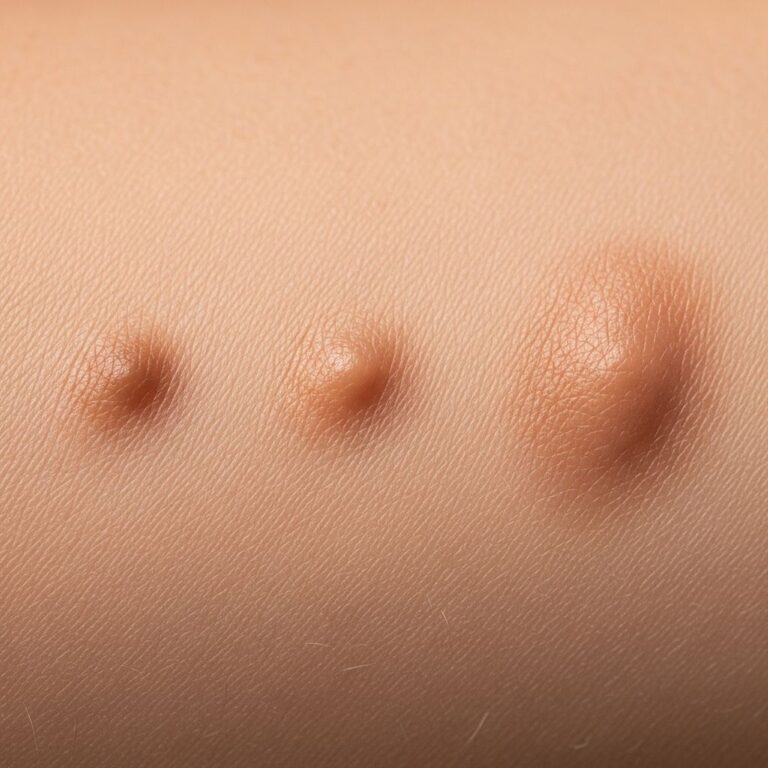
Epidermoid cysts present as firm, mobile, subcutaneous nodules ranging from 0.5 cm to several centimeters in diameter. A key diagnostic feature is the
central punctum
, a small blackhead-like opening representing the vestigial connection to the epidermis. Common locations include the face, neck, chest, back, scrotum, and genitals.- Slow growth over months to years
- Non-tender unless ruptured or infected
- Smooth, dome-shaped surface
- May transilluminate if large and superficial
When intact, cysts cause no symptoms beyond cosmetic concern. Rupture triggers an intense inflammatory response due to keratin leakage into the dermis, leading to redness, tenderness, swelling, and pus-like discharge. In rare cases, especially intracranial variants, expansion can compress adjacent structures causing headaches, neurological deficits, or seizures.
Symptoms of Inflammation or Infection
| Symptom | Description |
|---|---|
| Skin redness | Erythema surrounding the cyst |
| Tenderness | Pain on palpation |
| Warmth | Localized heat from inflammation |
| Discharge | Grayish-white, foul-smelling keratinous material |
Aetiology / Pathophysiology
The primary mechanism involves
traumatic implantation
of epidermal cells into the dermis, often from skin injury, surgical procedures, or acne. Blocked hair follicles trap epidermal cells, leading to cyst formation. Additional factors include:- UV light exposure
- Human papillomavirus (HPV) infection
- Developmental occlusion of follicular infundibula
Histopathologically, the cyst wall is lined by stratified squamous epithelium resembling epidermal infundibulum, with a granular layer producing keratohyalin granules. Keratin accumulates in laminated layers (onion-skin pattern) without cholesterol or cellular debris seen in dermoid cysts. Rupture releases keratin, eliciting a foreign body giant cell reaction and granulomatous inflammation.
In Gardner syndrome, multiple epidermoid cysts signal underlying genetic predisposition to colorectal cancer, warranting genetic evaluation.
Histopathology
Microscopic examination reveals a
cyst in the dermis
lined by thin, stratified squamous epithelium without rete ridges or adnexal appendages. The lumen containslaminated orthokeratotic keratin
with a granular layer present. If ruptured, surrounding dermis shows granulation tissue, multinucleated giant cells, and cholesterol clefts from keratin breakdown.- Epithelium: 2-3 layers thick, similar to infundibular epidermis
- Content: Acellular keratin flakes
- No skin appendages within wall
- Inflammation if disrupted
Differential histopathologic features distinguish from pilar cysts (trichilemmal keratinization) or vellus hair cysts.
Diagnosis
Diagnosis is primarily
clinical
, based on history and examination revealing a punctum-bearing nodule. No routine imaging or labs needed. Ultrasound may confirm cystic nature if atypical.Biopsy indicated for rapid growth, atypical location, or suspicion of malignancy (e.g., basal cell carcinoma arising in cyst). Incisional biopsy risks rupture and inflammation. Questions for providers include onset, trauma history, family cysts, and symptoms.
Differential Diagnosis
| Condition | Key Distinguishing Features |
|---|---|
| Lipoma | Softer, no punctum, subcutaneous fat |
| Dermoid cyst | Contains adnexa, congenital |
| Pilar cyst | Scalp, abrupt keratinization |
| Furuncle | Acute infection, no chronic punctum |
| Branchial cleft cyst | Neck, lateral, congenital |
Treatment / Management
Observation suffices for asymptomatic cysts.
Surgical excision
is curative, including cyst wall to prevent recurrence. Perform after inflammation subsides; use punch or elliptical incision through punctum.Infected cysts require incision and drainage (I&D), antibiotics, and delayed excision. Intralesional steroids reduce inflammation. Avoid squeezing to prevent scarring.
For recurrent or multiple cysts in Gardner syndrome, refer to genetics. Complete removal challenging in intracranial cases due to adhesions.
Prognosis and Complications
Excellent post-excision, with low recurrence if wall fully removed. Complications include infection (5-10%), scarring, and rare malignant transformation. Inflamed cysts mimic abscesses, delaying diagnosis.
Frequently Asked Questions (FAQs)
Q: Are epidermoid cysts cancerous?
A: No, they are benign, but rapid change warrants biopsy to exclude rare squamous cell carcinoma.
Q: Can epidermoid cysts resolve without treatment?
A: Rarely; they persist or inflame without intervention.
Q: What causes the foul smell from epidermoid cysts?
A: Bacterial decomposition of keratin after rupture.
Q: Is surgery the only treatment?
A: For cure, yes; conservative management for small asymptomatic lesions.
Q: Do epidermoid cysts run in families?
A: Common but familial clusters suggest Gardner syndrome evaluation.
Prevention
Avoid skin trauma and prompt acne treatment minimize risk. No proven preventive measures exist.
References
- Epidermoid cyst: MedlinePlus Medical Encyclopedia — MedlinePlus (National Library of Medicine). 2023. https://medlineplus.gov/ency/article/000842.htm
- Epidermoid Cyst Signs, Symptoms, Removal & Surgery — Pacific Neuroscience Institute. 2024. https://www.pacificneuroscienceinstitute.org/brain-tumor/conditions/epidermoid-cyst/
- Epidermoid Cyst – StatPearls — NCBI Bookshelf (National Center for Biotechnology Information). 2023-10-29. https://www.ncbi.nlm.nih.gov/books/NBK499974/
- Epidermoid Cysts of the Skin — UMass Memorial Health. 2024. https://www.ummhealth.org/health-library/epidermoid-cysts-of-the-skin
- Epidermal Inclusion Cysts (Sebaceous Cysts): Treatment & Causes — Cleveland Clinic. 2023-08-17. https://my.clevelandclinic.org/health/diseases/14165-sebaceous-cysts
- Epidermoid cysts – Diagnosis and treatment — Mayo Clinic. 2023-11-01. https://www.mayoclinic.org/diseases-conditions/epidermoid-cysts/diagnosis-treatment/drc-20352706
Similar Articles


Read full bio of medha deb