Epidermoid Cysts: Causes, Symptoms, Treatment Options
Discover causes, symptoms, diagnosis, and treatments for epidermoid cysts to manage these common skin lumps effectively.

Epidermoid cysts, often misnamed as sebaceous cysts, represent one of the most frequent benign skin conditions encountered in clinical practice. These slow-growing lumps form beneath the skin’s surface, typically containing a thick, protein-rich substance known as keratin. While generally harmless, they can become bothersome due to their visibility, discomfort, or potential for inflammation.
Understanding the Nature of Epidermoid Cysts
At their core, epidermoid cysts originate from the epidermis, the outermost layer of skin. They develop when surface skin cells multiply abnormally deep within the dermal layer instead of shedding naturally. This leads to the formation of a sac-like structure lined with squamous epithelial cells that continuously produce keratin, filling the cyst over time. Unlike true sebaceous cysts, which involve oil glands, epidermoid cysts do not produce sebum but rather a cheesy, waxy material composed primarily of keratin debris.
These cysts are encapsulated nodules, distinguishing them from other skin irregularities. They grow gradually, often reaching sizes from a few millimeters to several centimeters. Most remain stable, but external factors can trigger growth or complications.
Primary Causes and Formation Mechanisms
The exact trigger for epidermoid cyst development varies, but several well-documented mechanisms contribute. The most common pathway involves blockage of a hair follicle’s infundibulum, the upper portion where the follicle opens to the skin surface. This occlusion traps epidermal cells and keratin, prompting cyst formation.
- Follicular blockage: Often linked to acne or comedones, where dead skin and debris plug the pore.
- Skin trauma: Injuries like cuts, abrasions, or surgical incisions can implant epidermal cells into deeper layers.
- Sun damage: Chronic ultraviolet exposure, as seen in conditions like Favre-Racouchot syndrome, promotes cyst development in sun-exposed areas.
Rarely, genetic syndromes play a role. Conditions such as Gardner syndrome (associated with familial adenomatous polyposis) or Gorlin syndrome increase the likelihood of multiple cysts, particularly if they appear prepubertally or in atypical numbers/locations. Certain medications, including BRAF inhibitors, imiquimod, and cyclosporine, have also been implicated in cyst proliferation.
| Risk Factor | Description | Prevalence |
|---|---|---|
| Post-puberty age | Most common in adults 20-60 years | High |
| Skin injury | Trauma displaces epidermal cells | Moderate |
| Genetic syndromes | Gardner or Gorlin syndrome | Rare |
| Medications | BRAF inhibitors, etc. | Uncommon |
Common Locations and Demographic Patterns
Epidermoid cysts can emerge anywhere on the body but favor areas rich in hair follicles and sebaceous glands. Frequently affected sites include the face, neck, trunk, behind the ears, chest, back, scrotum, and genitals. Non-hair-bearing regions like palms, soles, or buttocks may develop cysts following trauma-induced epidermal implantation.
Demographically, they are more prevalent in men than women and rarely occur before puberty. Anyone can develop them, but individuals with a history of skin trauma or those in high-risk professions (e.g., manual laborers) face elevated chances.
Recognizing Symptoms and Signs
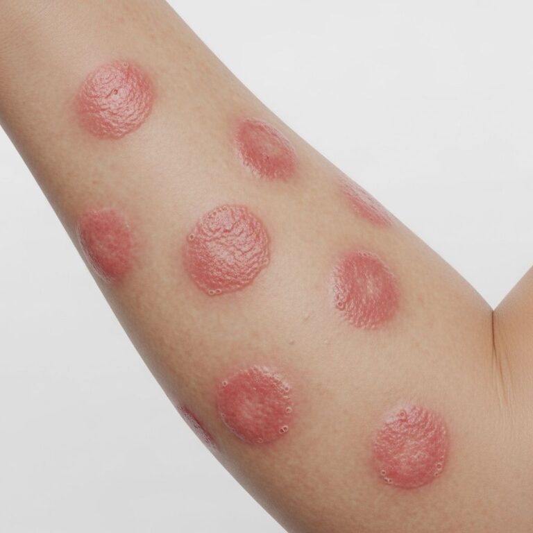
In their quiescent state, epidermoid cysts are typically asymptomatic, presenting as small, firm, dome-shaped nodules beneath the skin. They feel movable, rubbery, and may have a central punctum—a tiny blackhead-like opening from which keratin can occasionally express.
Symptoms arise primarily from complications:
- Inflammation: Redness, swelling, warmth, and tenderness if the cyst wall ruptures, spilling keratin into surrounding tissues.
- Infection: Pus drainage, fever, and severe pain from bacterial entry, often Staphylococcus aureus or Streptococcus.
- Growth: Enlargement causing cosmetic concerns or pressure on adjacent structures.
A ruptured cyst elicits a robust inflammatory response, mimicking an abscess. The overlying skin may thin, appearing shiny or translucent, with a foul-smelling discharge if infected.
Diagnostic Approaches
Diagnosis is primarily clinical, relying on visual inspection and palpation by a dermatologist. Key features include the cyst’s mobility, central punctum, and lack of adherence to deeper tissues. No routine imaging or lab tests are needed for straightforward cases.
Differential diagnoses encompass lipomas, dermoid cysts, hidradenomas, and rarely, basal cell carcinomas or squamous cell carcinomas (though malignant transformation is exceedingly rare). If uncertainty persists—especially for rapidly growing or atypical cysts—fine-needle aspiration, ultrasound, or biopsy may confirm the keratinous content and rule out malignancy.
Treatment Strategies and Options
Observation suffices for small, asymptomatic cysts. Intervention is warranted for painful, infected, recurrent, or cosmetically objectionable lesions.
Non-Surgical Methods
- Incision and drainage (I&D): Quick relief for inflamed cysts by evacuating contents. However, this risks recurrence without cyst wall removal.
- Intralesional steroids: Injections to shrink inflamed cysts temporarily.
- Antibiotics: Oral or topical for infected cases, targeting common pathogens.
Surgical Excision
The gold standard for definitive treatment involves complete surgical excision under local anesthesia. The entire cyst wall must be removed to prevent regrowth. Minimal excision techniques minimize scarring. Post-operative care includes wound hygiene to avert infection.
Recurrence rates are low (<5%) with proper technique but rise if the sac fragments during removal.
Potential Complications and Prognosis
Complications are infrequent but include infection, scarring from rupture or surgery, and rare malignant transformation (e.g., squamous cell carcinoma within the cyst). Prognosis is excellent following appropriate management; most patients achieve full resolution without sequelae.
Multiple cysts may signal underlying syndromes, necessitating genetic evaluation.
Prevention and Daily Management Tips
While not entirely preventable, minimizing skin trauma, prompt acne treatment, and sun protection reduce risks. Avoid squeezing cysts, as this promotes inflammation and scarring. Maintain gentle skincare routines without aggressive scrubbing.
Frequently Asked Questions (FAQs)
Are epidermoid cysts cancerous?
No, they are benign. Malignant changes are exceptionally rare.
Can I pop an epidermoid cyst at home?
No, this heightens infection and recurrence risks. Seek professional care.
How long does recovery take after removal?
Typically 1-2 weeks for healing, with stitches removed in 7-14 days.
Do epidermoid cysts go away on their own?
Rarely; they persist unless treated.
Are they contagious?
No, they do not spread person-to-person.
Living with Epidermoid Cysts
For many, these cysts pose minimal disruption. Regular dermatologic check-ups aid early detection of changes. Expanded discussion on cyst biology reveals their derivation from follicular infundibulum proliferation, where stratified squamous epithelium lines the cyst, perpetuating keratin accumulation. Pathophysiologically, rupture induces granulomatous inflammation due to keratin’s foreign-body reaction. Recent studies link HPV and UV exposure to pathogenesis, broadening etiological understanding.
In clinical cohorts, prevalence underscores their ubiquity: up to 40% of excised skin cysts are epidermoid. Men exhibit higher scrotal involvement, while facial cysts dominate cosmetically motivated excisions. Advanced imaging like MRI delineates intracranial variants, though cutaneous forms predominate.
Treatment evolution favors minimally invasive approaches, such as punch biopsy excision for small lesions, preserving aesthetics. Post-excision histology confirms diagnosis, revealing onion-skin keratin lamellae. Patient education emphasizes hygiene sans over-cleansing, debunking hygiene causation myths.
Genetic counseling benefits syndromic cases; Gardner syndrome screening includes colonoscopy for polyposis. Pharmacovigilance identifies drug-induced cysts, prompting therapy adjustments. Overall, epidermoid cysts exemplify manageable dermatoses, blending conservative and interventional strategies for optimal outcomes.
References
- Epidermoid Cyst: Causes, Diagnosis, and Treatments — Healthline. 2023. https://www.healthline.com/health/epidermoid-cysts
- Epidermoid Cyst — StatPearls, NCBI Bookshelf, NIH. 2023-10-01. https://www.ncbi.nlm.nih.gov/books/NBK499974/
- Epidermoid cysts – Symptoms and causes — Mayo Clinic. 2023. https://www.mayoclinic.org/diseases-conditions/epidermoid-cysts/symptoms-causes/syc-20352701
- Epidermal Inclusion Cysts (Sebaceous Cysts): Treatment & Causes — Cleveland Clinic. 2023. https://my.clevelandclinic.org/health/diseases/14165-sebaceous-cysts
- Epidermoid cyst — DermNet NZ. 2023. https://dermnetnz.org/topics/epidermoid-cyst
Similar Articles


Read full bio of Sneha Tete