Epidermolytic Ichthyosis: Causes, Symptoms & Management
Understanding epidermolytic ichthyosis: genetic causes, clinical features, and evidence-based symptom management strategies.

Epidermolytic Ichthyosis: Overview and Clinical Significance
Epidermolytic ichthyosis (EI) is a rare genetic skin disorder characterized by severe skin fragility, erythroderma, and abnormal scaling that typically manifests at birth. Previously known as epidermal hyperkeratosis and bullous congenital ichthyosiform erythroderma, this condition fundamentally impairs the skin’s barrier function, affecting the outermost epidermal layers. The disorder results from inherited or sporadic mutations in genes encoding structural proteins essential for skin integrity, leading to progressive skin complications throughout life. As the largest organ of the body, the skin serves multiple critical functions including providing a protective barrier, retaining moisture, regulating vitamin D production, and transmitting sensations. When these functions are compromised by epidermolytic ichthyosis, numerous complications can develop. Understanding this condition is essential for patients, caregivers, and healthcare providers to implement effective management strategies and improve quality of life.
Genetic Basis and Molecular Pathophysiology
Epidermolytic ichthyosis is caused by missense mutations in keratin genes, specifically keratin 1 (KRT1) or keratin 10 (KRT10). These mutations result in improper expression of keratin proteins, which are structural proteins specifically found in epithelial and epidermal cells. The malfunctioning proteins result in barrier abnormalities that lead to epidermal inflammation and the characteristic symptoms observed in the disease.
A significant clinical observation involves the relationship between mutation burden and disease severity. Children with mutations in some but not all of their cells tend to present with a less severe form of the condition. Conversely, individuals with mutations affecting a higher percentage of cells experience more pronounced symptoms and complications. This mosaic pattern of genetic expression explains the variable phenotypic presentations observed among patients with epidermolytic ichthyosis.
The inheritance pattern for epidermolytic ichthyosis is typically autosomal dominant, meaning affected individuals have a 50% chance of passing the condition to their offspring. However, sporadic mutations can also occur, accounting for cases in which neither parent carries the genetic variant.
Clinical Presentation and Symptoms
The clinical manifestations of epidermolytic ichthyosis vary depending on disease severity, patient age, and affected body regions. However, several characteristic features define the condition across patient populations.
Neonatal and Early Presentation
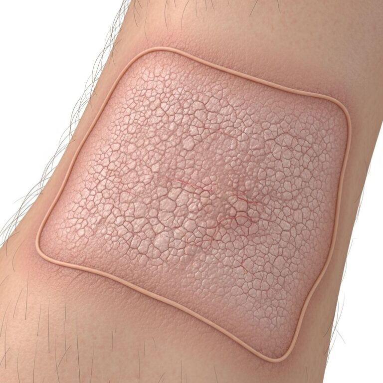
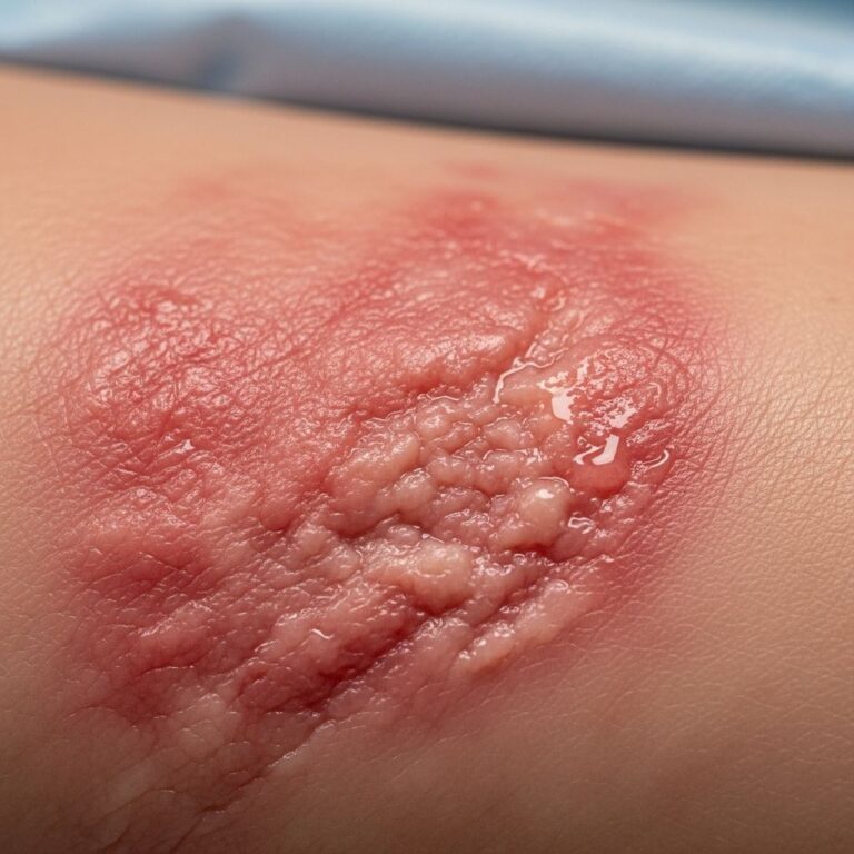
Infants born with epidermolytic ichthyosis present with distinctly abnormal skin appearance at birth. Visibly red areas of skin (erythroderma) and blistering are the most common signs when an infant is born with EI. These initial manifestations reflect the underlying defect in skin structure and barrier function. Scales tend to develop not long after birth and often form in parallel rows. The skin appears extremely fragile and hypersensitive, with increased susceptibility to damage from minor trauma.
Infants with severe presentations may require immediate specialized care. If symptoms are evident at birth, the newborn is typically transferred to the neonatal intensive care unit (ICU) for proper monitoring and management of potential complications.
Systemic Symptoms in Children and Adults
Beyond the visible skin changes, epidermolytic ichthyosis affects multiple physiological systems:
- Anhidrosis and Heat Intolerance: Difficulty sweating occurs because thick layers of skin clog the sweat glands, resulting in reduced thermoregulation and heat sensitivity
- Pruritus: Persistent itching is a hallmark symptom that can severely impact quality of life and lead to excessive scratching, causing secondary skin wounds
- Xerosis: Abnormal dryness of the skin characterizes the condition, requiring intensive moisturizing regimens
- Fissuring and Skin Fragility: The skin cracks easily and frequently becomes infected, with decreased joint mobility in severe cases
- Ocular Manifestations: Dry eyes occur due to compromised lacrimal function and epithelial changes
- Hair and Nail Abnormalities: Thin or fragile hair results from buildup of skin scales that block hair follicles, while nail abnormalities may also be present
- Otologic Complications: Issues with hearing can develop from buildup of skin scales in the ears
- Palmoplantar Keratoderma: Thickened skin on the palms and soles may develop and can become so severe as to limit movement and hand function
Histological Features
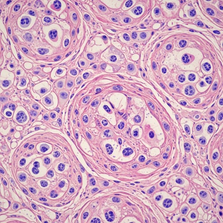
Under microscopic examination, epidermolytic ichthyosis exhibits distinctive pathological findings. The skin demonstrates mid-epidermal splitting and skin thickening (hyperkeratosis), with acanthosis and a prominent granular layer evident on histological analysis. Electron microscopic examination reveals structural alterations of tonofilaments in keratinocytes of the granular layer.
Associated Complications
Complications of epidermolytic ichthyosis are due to the impaired skin barrier function, which renders the epidermis vulnerable to multiple threats. Understanding and preventing these complications is central to disease management:
- Bacterial and Secondary Skin Infections: Compromised barrier function and skin ulcers create portals for bacterial colonization. Skin infections represent one of the most common and serious complications requiring aggressive preventive and therapeutic measures
- Dehydration: Excessive transepidermal water loss through the damaged skin barrier can lead to systemic dehydration, particularly in infants
- Heat Intolerance: The inability to dissipate body heat effectively through impaired sweat gland function places patients at risk for hyperthermia and related complications
- Psychological Distress: The cosmetic issues and sometimes unpleasant odor associated with epidermolytic ichthyosis can lead to significant stress and psychological distress
- Mobility Limitations: Severe palmoplantar keratoderma can restrict hand function and mobility
- Structural Abnormalities: Reports exist of palmoplantar contractures developing in some patients
- Neoplastic Risk: Epidermolytic ichthyosis is associated with atypical naevi, warranting careful dermatological monitoring
Diagnosis and Differential Considerations
Diagnosis of epidermolytic ichthyosis is based on clinical presentation, histological examination of skin biopsies, and molecular genetic analysis. The combination of congenital erythroderma, blistering, and characteristic mid-epidermal splitting on histology strongly suggests the diagnosis.
Molecular analysis identifying mutations in KRT1 or KRT10 genes confirms the diagnosis. Several related conditions exhibit overlapping features but result from mutations in different genes. Erythrokeratodermia variabilis (EKV) and keratitis-ichthyosis-deafness (KID) like disorders are caused by variants in genes coding for gap junction proteins (GJB3, GJB4, and GJB2 respectively) and present with distinct clinical features.
Superficial epidermolytic ichthyosis (SEI), a related disorder, is caused by mutations in the KRT2 gene and typically presents with less severe symptoms, including superficial blisters and erosions at birth.
Management and Treatment Approach
There is no cure for epidermolytic ichthyosis, and treatment focuses on symptom management and complication prevention. The main goal of treating epidermolytic ichthyosis is to ease the presenting symptoms, which can be challenging as it typically requires a combination of treatments tailored to individual needs.
Foundational Skin Care Routine
The cornerstone of epidermolytic ichthyosis management involves a rigorous daily skin care routine:
- Frequent Bathing and Cleansing: Bathing or showering frequently using a mild, soap-free cleanser softens the skin and removes layers of thickened skin. Many patients find baths with salt or sodium bicarbonate, as well as bleach, beneficial as these solutions help to descale and prevent bacterial overgrowth
- Mechanical Debridement: Rubbing the skin lightly with a loofah, rough-textured sponge, or pumice stone helps remove thickened skin. This gentle mechanical approach prevents excessive damage to fragile underlying tissue
- Emollient Application: Applying moisturizers to the skin immediately after bathing and throughout the day reduces skin dryness. Tools like pumice stone and skin products containing lanolin, urea, propylene glycol, or alpha hydroxy acids may be helpful. Application of a barrier repair formula containing ceramides or cholesterol may be used to relieve and protect vulnerable skin
Pharmacological Treatments
Several medication classes are employed in epidermolytic ichthyosis management:
- Topical Keratolytics: These medications help shed the damaged outer layer of skin. Products containing salicylic acid are commonly used. However, these agents can leave the fragile epidermis exposed, necessitating concurrent use of protective emollients
- Topical Emollients: These soothe and soften the skin, protecting the exposed epidermis from environmental irritants and transepidermal water loss
- Retinoids: Topical or oral retinoids may greatly lessen symptoms by preventing hyperkeratinization and reducing superinfection in severe cases. However, retinoids present a paradox: they can reduce skin scaling significantly, but if discontinued, the scales return. Long-term use of retinoids can cause serious adverse effects, so doctors prescribe them only when symptoms are very severe. Oral retinoids must be used with caution because of their side effects and their effect in increasing skin fragility
- Antiseptic Skin Wash: An antiseptic skin wash helps remove bacteria from the skin to reduce infection risk. It is recommended that patients wash with antiseptic soap 2-3 times per week
- Antibiotics: Treatment with antibiotics may be needed when a skin infection is suspected, either topical or systemic depending on infection severity and extent
Specialized Care in Severe Cases
Severe presentations require intensive management. Developing and sticking with a daily routine aimed at keeping the skin moist is important, as this approach helps remove dead and thickened areas of skin and controls skin itching and cracking. In neonatal cases, specialized care in an intensive care setting ensures proper monitoring for complications including infection, dehydration, and thermoregulatory disturbances.
Long-term Outlook and Quality of Life Considerations
While epidermolytic ichthyosis is a lifelong condition without cure, many patients achieve reasonable symptom control through diligent skin care and appropriate medical management. The severity can vary significantly between individuals and even fluctuate in the same individual over time. Some patients experience improvement in symptoms during childhood, while others face persistent or worsening manifestations.
The psychological and social impact of epidermolytic ichthyosis should not be underestimated. The visible skin changes, odor, and ongoing treatment requirements can affect emotional well-being and social integration. Multidisciplinary approaches incorporating dermatological, psychological, and social support optimize overall patient outcomes.
Genetic Counseling and Family Planning
Affected families should be offered genetic counseling to understand inheritance patterns and reproductive risks. For autosomal dominant epidermolytic ichthyosis, the risk for an affected parent to have an affected child is 50%. Prenatal diagnosis may be available through molecular genetic testing in some cases, enabling informed reproductive decision-making.
Frequently Asked Questions
Q: Is epidermolytic ichthyosis contagious?
A: No, epidermolytic ichthyosis is not contagious. It is a genetic disorder caused by inherited mutations in keratin genes that affect skin structure and function.
Q: Can epidermolytic ichthyosis be cured?
A: Currently, there is no cure for epidermolytic ichthyosis. Treatment focuses on managing symptoms, preventing complications, and improving quality of life through skin care regimens and medications.
Q: What is the prognosis for children born with epidermolytic ichthyosis?
A: The prognosis varies depending on disease severity and mutation burden. Many children with appropriate management experience reasonable symptom control, though the condition typically persists into adulthood. Early neonatal care and intensive management significantly improve outcomes.
Q: Are there any new treatments under development for epidermolytic ichthyosis?
A: Research into novel therapeutic approaches is ongoing. Gene therapy and other advanced treatment modalities show promise in preclinical and early clinical studies, though these remain investigational at present.
Q: How often should patients with epidermolytic ichthyosis bathe?
A: Frequent bathing using mild, soap-free cleansers is recommended. Many patients benefit from daily or near-daily bathing to soften skin and remove thickened scales, followed immediately by application of emollients to lock in moisture.
Q: Can epidermolytic ichthyosis improve with age?
A: Some patients experience improvement in symptoms during childhood, though the condition remains chronic. Symptom severity can vary significantly between individuals and may fluctuate over time with proper management.
References
- Epidermolytic Ichthyosis Symptoms and Treatment — Hines Dermatology Associates. https://www.hinesdermatologyassociates.com/blog/epidermolytic-ichthyosis-symptoms-and-treatment
- Epidermolytic Ichthyosis – Symptoms, Causes, Treatment — National Organization for Rare Disorders (NORD). https://rarediseases.org/rare-diseases/epidermolytic-ichthyosis/
- Epidermolytic Ichthyosis — First Skin Foundation. https://www.firstskinfoundation.org/types-of-ichthyosis/epidermolytic-ichthyosis
- Epidermolytic Ichthyosis — DermNet. https://dermnetnz.org/topics/epidermolytic-ichthyosis
- Superficial Epidermolytic Ichthyosis — Orphanet. https://www.orpha.net/en/disease/detail/455
- Epidermolytic Hyperkeratosis — StatPearls, National Center for Biotechnology Information (NCBI). https://www.ncbi.nlm.nih.gov/books/NBK544323/
Similar Articles


Read full bio of medha deb