Episcleritis and Scleritis: Causes and Treatment
Understanding eye inflammation: Learn to distinguish episcleritis from scleritis and treatment options.

Episcleritis and Scleritis: Understanding Eye Inflammation
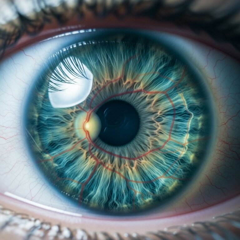
The eye is a complex organ composed of multiple layers, each serving a critical function in vision. When inflammation affects the tissues surrounding and supporting the eye, it can cause discomfort and affect vision quality. Two conditions that commonly affect these tissues are episcleritis and scleritis. While both conditions involve inflammation of the eye and present with similar initial symptoms such as redness, they are distinct conditions with different severities, treatment approaches, and potential complications. Understanding the differences between these conditions is essential for proper diagnosis and management.
What is Episcleritis?
Episcleritis is an inflammation of the episclera, the thin layer of tissue between the conjunctiva (the clear membrane covering the white of the eye) and the sclera (the white part of the eye). The episclera is composed of loose connective tissue with a rich vascular supply derived from the anterior ciliary arteries, which are branches of the ophthalmic artery. This condition is characterized by non-granulomatous inflammation of the episcleral vascular network, leading to edema and dilation of the radially oriented superficial blood vessels.
Episcleritis is a relatively mild condition that usually resolves on its own within a few weeks. It is more common than scleritis and typically does not pose a serious threat to vision. The condition can occur in one or both eyes and may be associated with systemic autoimmune diseases, collagen vascular diseases, and certain infections, though it is most often idiopathic (occurring without a known cause).
What is Scleritis?
Scleritis is a more severe and potentially serious inflammation of the sclera itself, the deeper white part of the eye. Unlike episcleritis, which affects only the superficial episcleral layer, scleritis involves the sclera and can affect deeper eye structures. Scleritis is a serious condition that can cause significant eye pain, redness, and vision problems. It is often associated with underlying autoimmune diseases and requires medical treatment. The condition is not as common as episcleritis but is far more serious because the inflamed vessels are deeper in the eye.
Types of Episcleritis
Episcleritis presents in two main forms, each with distinct characteristics:
- Diffuse Episcleritis: This is the most common type and typically causes the surface of the eye to become evenly red, sometimes across the entire eye and sometimes in a wedge shape. Diffuse episcleritis is usually idiopathic and presents with benign, mild inflammation that resolves within days to weeks.
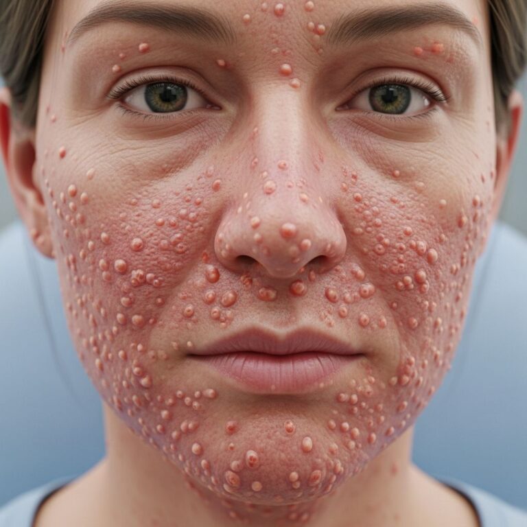
- Nodular Episcleritis: This less common type comes on more slowly and causes inflamed, swollen bumps or nodules in the episclera. Nodular episcleritis is more painful than diffuse episcleritis, lasts longer, and is more likely to be associated with an underlying inflammatory or systemic autoimmune disorder in up to one-third of cases.
Types of Scleritis
Scleritis is classified into several types based on the location and severity of inflammation:
- Anterior Scleritis: This involves inflammation of the sclera anterior to the extraocular muscle insertion and accounts for the majority of scleritis cases. Anterior scleritis has three variants:
Diffuse Anterior Scleritis: The most common variant of anterior scleritis and also the most treatable, presenting with widespread inflammation across the anterior sclera.
Nodular Anterior Scleritis: Presents with tender, painful nodules on the scleral surface.
Necrotizing Anterior Scleritis: The most severe and destructive form, characterized by significant scleral necrosis and potential vision loss.
- Posterior Scleritis: This rarer form involves inflammation of the sclera posterior to the extraocular muscle insertion. It is difficult to detect due to its more posterior location and is associated with serious complications including retinal detachment, angle-closure glaucoma, and vision loss.
Symptoms of Episcleritis
Episcleritis presents with relatively mild symptoms that distinguish it from scleritis:
- Redness of the eye, typically localized to a specific area or diffuse across the globe
- Mild discomfort or irritation of the eye
- A feeling of grittiness
- Mild eye discomfort with acute onset
- Possible epiphora (excessive tearing), though this is not always present
- Photophobia may occur in some cases
- Normal or preserved visual acuity—vision loss does not occur
Importantly, episcleritis typically affects only one eye and does not usually cause vision problems. The discomfort is generally mild and does not significantly impact daily activities.
Symptoms of Scleritis
Scleritis presents with much more severe symptoms that require urgent medical attention:
- Severe eye pain: Scleritis is characterized by significant and often intense pain that is disproportionate to clinical findings. The pain is typically described as a deep, boring ache that may radiate to the face, cheek, and jaw.
- Pain with eye movement, often exacerbated by movement
- Pain that is typically worse at night and often interferes with sleep
- Redness and swelling of the eye, which tends to develop more slowly than in episcleritis
- Sensitivity to light (photophobia)
- Blurry vision and potential reduced visual acuity
- Excessive tearing (lacrimation)
- Conjunctival injection (redness of the conjunctiva)
The pain associated with scleritis can be significantly more severe than that of episcleritis, often causing considerable distress and requiring immediate evaluation.
Key Differences Between Episcleritis and Scleritis
| Feature | Episcleritis | Scleritis |
|---|---|---|
| Severity | Mild, self-limited condition | Serious, potentially sight-threatening |
| Pain Level | Mild discomfort or irritation | Severe, deep, boring pain |
| Vision Loss | No vision loss | Risk of vision loss |
| Photophobia | No photophobia | Photophobia present |
| Vessel Appearance | Superficial, radial pattern of vessels | Deep, fixed, criss-crossed vessel pattern |
| Scleral Hue | Reddish hue | Red-purple or violet hue |
| Duration | Days to weeks | Longer duration, requires treatment |
| Treatment | Self-limited, may not require treatment | Requires ophthalmology care and medication |
Diagnostic Methods
Proper diagnosis is crucial to distinguish episcleritis from scleritis and guide appropriate treatment. Several diagnostic techniques are employed:
Clinical Examination
The initial evaluation involves careful observation of the eye’s appearance and symptoms. Healthcare providers assess the location and pattern of redness, the presence of nodules, and the patient’s pain level.
Phenylephrine Test
Phenylephrine ophthalmic drops are a key diagnostic tool. When applied to the eye, these drops cause vasculature to blanch (whiten). In episcleritis, the superficial episcleral vessels will blanch completely with phenylephrine drops. In contrast, the deeper scleral vessels in scleritis do not blanch with phenylephrine because the medication does not reach deeper vessels. This simple test helps differentiate between the two conditions.
Cotton Swab Test
Another useful diagnostic technique involves gently moving the eye’s surface with a cotton swab. In episcleritis, the superficial vessels are mobile and can be easily moved. In scleritis, the deeper vessels are firmly attached to the sclera and cannot be moved, remaining fixed in position.
Slit Lamp Examination
A slit lamp is a microscope that allows detailed examination of the eye’s structures. It can be used to distinguish episcleritis from nodular or diffuse scleritis and to look for the characteristic blue-violet hue associated with scleritis. This examination may also reveal scleral thinning, chemosis (swelling), anterior chamber cells and flare, and other signs of deeper inflammation.
Causes and Associated Conditions
Episcleritis Causes
Most cases of episcleritis, particularly diffuse episcleritis, are idiopathic (no identifiable cause). However, episcleritis can be associated with systemic conditions, particularly autoimmune and collagen vascular diseases. Nodular episcleritis is more likely to be associated with underlying systemic disease. Associated conditions may include various autoimmune disorders and some infections.
Scleritis Causes
Scleritis is frequently associated with systemic disease and often indicates an underlying condition that requires investigation. The condition is commonly linked to autoimmune and rheumatologic disorders. Women between the ages of 30 and 50 with rheumatologic disorders are at particular risk. Additionally, corneal abrasions can lead to scleritis development. The presence of herpes zoster virus (HZV) is associated with particularly severe pain in scleritis cases.
Treatment of Episcleritis
Episcleritis is typically self-limited and often does not require acute treatment. Most cases resolve spontaneously within days to weeks without intervention. However, symptomatic relief may be provided through:
- Observation and watchful waiting, allowing the condition to resolve naturally
- Artificial tears to provide comfort and lubrication
- Cool compresses to reduce inflammation and discomfort
- Non-steroidal anti-inflammatory drugs (NSAIDs) if pain management is needed
If the condition persists or causes significant discomfort, topical corticosteroids may be considered, though these are generally not necessary for straightforward episcleritis.
Treatment of Scleritis
Unlike episcleritis, scleritis requires prompt and aggressive medical treatment to prevent serious complications and vision loss. Treatment typically involves:
- Immediate ophthalmology consultation: Scleritis should always be referred to an eye specialist for proper management.
- Systemic corticosteroids: Oral corticosteroids are commonly prescribed to control inflammation and manage pain.
- Topical medications: Topical corticosteroids and antibiotics may be used as adjunctive therapy.
- Investigation for underlying disease: Because scleritis is often associated with systemic disease, diagnostic workup to identify underlying conditions is essential.
- Immunosuppressive therapy: For severe cases or those associated with serious systemic disease, additional immunosuppressive medications may be necessary.
- Treatment of underlying conditions: Management of any associated autoimmune or rheumatologic disease is crucial.
When to Seek Medical Care
While episcleritis is generally benign and self-resolving, certain symptoms warrant professional evaluation. Patients should seek medical attention if:
- Eye pain is severe or progressive
- Vision changes occur
- Symptoms persist beyond a few weeks
- Photophobia (sensitivity to light) develops
- The condition affects both eyes simultaneously
- Accompanying systemic symptoms are present
Scleritis always requires immediate medical evaluation and should never be managed without professional ophthalmologic care.
Prognosis and Complications
Episcleritis Prognosis
The prognosis for episcleritis is excellent. Most cases resolve completely within days to weeks without any lasting effects or vision loss. Recurrence can occur, but this does not typically cause any long-term damage to the eye.
Scleritis Complications
Scleritis carries significant risk for serious complications if not treated appropriately. Potential complications include vision loss, scleral thinning and staphyloma (outward bulging of the sclera), corneal involvement, and conditions such as retinal detachment and angle-closure glaucoma in posterior scleritis.
Frequently Asked Questions
Q: Can episcleritis return after it resolves?
A: Yes, episcleritis can recur, though this is not always the case. Recurrent episodes typically follow the same self-limited course as the initial episode and do not cause permanent damage.
Q: Is episcleritis contagious?
A: No, episcleritis is not contagious. It is an inflammatory condition, not an infection, and cannot be transmitted to other people.
Q: Can episcleritis affect vision?
A: No, episcleritis does not cause vision loss. Vision remains normal despite the presence of eye redness and discomfort.
Q: How is scleritis different from episcleritis?
A: Scleritis is a much more serious condition affecting deeper eye structures and causes severe pain, potential vision loss, and requires medical treatment. Episcleritis is mild, self-limiting, and does not cause vision problems or severe pain.
Q: Should I see a doctor for episcleritis?
A: While episcleritis often resolves without treatment, seeing a doctor can help confirm the diagnosis and rule out scleritis. Seek medical care if symptoms persist beyond a few weeks or if vision changes occur.
Q: What should I do if I suspect I have scleritis?
A: If you suspect scleritis based on severe eye pain and other symptoms, seek immediate medical attention from an ophthalmologist. Do not delay treatment, as scleritis requires prompt medical intervention.
References
- Episcleritis and Scleritis – Horizon Eye Physicians — Horizon Eye Physicians. Accessed January 2026. https://horizoneye.org/conditions/episcleritis-and-scleritis-2/
- Scleritis and Episcleritis — Taming the SRU. Accessed January 2026. https://www.tamingthesru.com/blog/bread-butter-em/scleritis-and-episcleritis
- A Red Eye: Scleritis or Episcleritis? — Review of Optometry. Accessed January 2026. https://www.reviewofoptometry.com/article/ro1117-a-red-eye-scleritis-or-episcleritis
- Scleritis vs. Episcleritis — Yale Emergency Medicine. Accessed January 2026. https://medicine.yale.edu/emergencymed/education/resources/slit-lamp/eye-pain/scleritis-vs-episcleritis/
- Episcleritis – StatPearls — National Center for Biotechnology Information (NCBI), National Institutes of Health (NIH). Accessed January 2026. https://www.ncbi.nlm.nih.gov/books/NBK534796/
- Distinguishing Episcleritis from Scleritis in Optometric Practice — Review Education Group. Accessed January 2026. https://www.revieweducationgroup.com/ce/distinguishing-episcleritis-from-scleritis-in-optometric-practice
Similar Articles


Read full bio of medha deb