Erosive Pustular Dermatosis: Causes, Symptoms & Treatment
Understanding erosive pustular dermatosis: causes, clinical presentation, and evidence-based treatment options.

Erosive Pustular Dermatosis: A Comprehensive Overview
Erosive pustular dermatosis (EPD) is a rare but significant skin condition that predominantly affects elderly individuals, particularly those with fair skin. While the scalp remains the most commonly affected site, this condition can also manifest on the forehead, temples, and lower legs. The disease is characterized by the development of pustules, erosions, and crusts that can progress to permanent scarring and hair loss if left untreated. Understanding the etiology, clinical presentation, and management of EPD is crucial for healthcare providers to ensure accurate diagnosis and optimal patient outcomes.
Understanding the Cause and Risk Factors
The exact etiology of erosive pustular dermatosis remains unknown, yet research and clinical observations have identified several contributing factors. Sun damage and actinic injury appear to play a significant role in disease development, with cumulative ultraviolet exposure contributing to skin changes that predispose individuals to EPD. The condition typically emerges following minor trauma to the affected area, a phenomenon known as pathergy, which can include surgical procedures, minor injuries, or skin interventions.
Several specific triggers have been documented as precursors to EPD development:
- Physical trauma and surgical procedures
- Cryotherapy applications
- Topical chemotherapy agents
- Carbon dioxide laser therapy
- Imiquimod treatment
- Photodynamic therapy with topical methyl aminolevulinate
- Skin grafting procedures following nonmelanoma skin cancer surgery
- Herpes zoster infections
- Cochlear implant positioning
Beyond direct trauma, EPD has been associated with various systemic and autoimmune conditions, including rheumatoid arthritis, autoimmune hepatitis, Hashimoto thyroiditis, and Takayasu aortitis. Additionally, drug-induced cases have been reported with latanoprost, minoxidil, and epidermal growth factor receptor (EGFR) inhibitors such as gefitinib.
Environmental and procedural factors also contribute to disease risk. Treatments targeting actinic keratoses and androgenetic alopecia can trigger lesion development in susceptible individuals. When EPD affects the lower legs, venous stasis and edema may be involved in pathogenesis. Importantly, infection is not considered the primary cause, as lesions do not resolve with antibiotics alone, though secondary bacterial infection may occur in moist lesions.
Clinical Presentation and Symptoms
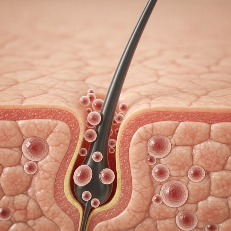
The clinical manifestations of erosive pustular dermatosis are distinctive and progressive. The condition typically initiates with tiny pustules on the scalp, forehead, or temples, which then evolve into characteristic lesions. Key clinical features include:
- Pustules: Small, pus-filled blisters that frequently rupture, leading to crust formation
- Erosions and Ulcers: Shallow, painful sores of varying sizes that may coalesce into larger affected areas
- Crusting: Thick yellow-brown or greenish crusts that form over pustules and erosions
- Inflammation: Marked erythema and swelling surrounding lesions
- Discomfort: Patients often report pain, burning sensations, or pruritus in affected areas
- Scarring Alopecia: When located on the scalp, progressive lesions can result in permanent hair loss and visible scarring
Upon removal of crusts, the underlying skin typically appears red and moist. Extensive disease can itself trigger scarring and significant baldness. The condition’s chronic nature means symptoms may persist or recur over extended periods, significantly impacting quality of life.
Diagnosis and Diagnostic Procedures
Clinical diagnosis of erosive pustular dermatosis is primarily based on characteristic clinical appearance combined with patient history and physical examination findings. Trichoscopy, the microscopic examination of hair and scalp structures, can provide additional diagnostic support. However, scalp biopsy is recommended to confirm diagnosis and assess histopathological features, though biopsy findings are not pathognomonic for the condition.
Histopathological examination typically reveals:
- Epidermal atrophy
- Focal erosions
- Lymphoplasmacytic infiltrate with or without foreign body giant cells
- Pilosebaceous atrophy
- Mixed inflammatory infiltrate containing neutrophils, lymphocytes, and plasma cells
Culture of bacterial swabs may reveal secondary bacterial colonization, though cultures are usually negative. Fungal and viral stains are not necessary as infections are not primary pathogens. This distinction is critical to avoid misdiagnosis as a bacterial or fungal infection. Gram-negative bacteria may occasionally be present in moist lesions but represent secondary contaminants rather than primary causative agents.
Complications of Erosive Pustular Dermatosis
The primary complications of EPD relate to its chronic inflammatory nature and progression:
- Scarring Alopecia: Permanent hair loss in affected scalp areas due to destruction of hair follicles
- Secondary Bacterial Infection: Moist lesions may become colonized with bacteria, requiring antibiotic treatment
- Permanent Disfigurement: Extensive scarring can result in visible cosmetic changes
- Psychological Impact: Chronic lesions and hair loss can affect patient psychological well-being and quality of life
- Treatment Challenges: Some cases prove refractory to standard therapies, necessitating prolonged or escalated treatment regimens
Treatment Approaches and Therapeutic Options
Although no universally recognized treatment guideline exists for EPD due to its rarity and unclear etiology, topical treatments represent the first-line approach. Therapy aims to reduce inflammation, heal erosions, halt scarring alopecia progression, and minimize permanent hair loss. Early intervention is crucial for optimal outcomes.
First-Line Treatments
High-potency topical corticosteroids remain the gold standard therapy, with demonstrated efficacy in the majority of documented cases. Agents such as clobetasol and betamethasone are effective in controlling the disorder and drying lesions. These should be applied once or twice daily to affected areas for several weeks, with repeated applications as necessary when recurrence occurs. Treatment duration typically ranges from weeks to months, with response varying among patients.
Topical tacrolimus 0.1% ointment has emerged as a viable alternative to corticosteroids, demonstrating comparable efficacy. This calcineurin inhibitor is particularly beneficial when long-term topical steroid application is required, helping to avoid complications associated with prolonged corticosteroid use.
Supportive Care and Wound Management
Essential to successful treatment is good local skin care, including gentle removal of crusts and daily dressing of affected areas. This supportive approach promotes direct healing and improved outcomes, delivering positive quality-of-life benefits for this otherwise recalcitrant condition.
Second-Line and Alternative Treatments
For patients with inadequate response to empiric treatment, additional therapeutic options include:
- Topical Retinoids: Oral or topical formulations to reduce inflammation and promote healing
- Topical Calcipotriol: Vitamin D analog useful in refractory cases
- Photodynamic Therapy (PDT): Effective in some patients, though controversially, PDT has induced disease in others; use requires careful consideration
- Dapsone: Oral or gel formulations for refractory disease
- Systemic Corticosteroids: Short courses for severe or rapidly progressing disease
- Doxycycline and Isotretinoin: Alternative systemic agents for difficult-to-treat cases
- Acitretin: Systemic retinoid for refractory erosive pustular dermatosis
- Oral Zinc Sulfate: Adjunctive therapy supporting skin healing
- Silicone Gel: Supportive wound care adjunct
Treatment of Secondary Infection
When secondary bacterial infection develops, oral anti-Staphylococcal antibiotics such as flucloxacillin or erythromycin should be initiated. This treatment addresses bacterial colonization of moist lesions without implying infection as a primary cause.
Clinical Considerations and Prognosis
Erosive pustular dermatosis is a chronic, recurring condition requiring extended therapy and careful long-term management. While response to treatment is generally good, efficacy varies significantly among patients. Relapses following corticosteroid discontinuation are common, necessitating maintenance therapy or rapid reinitiation when symptoms recur.
The resulting alopecia may or may not be reversible, depending on disease duration, severity, and treatment timing. Early intervention offers the best opportunity to minimize permanent hair loss and scarring. For patients with inadequate response to initial therapy, biopsy confirmation should be considered to ensure accurate diagnosis before escalating treatment intensity.
Pathophysiology and Disease Mechanisms
Current understanding suggests that four key elements contribute to EPD development: (1) initial trauma to the scalp, (2) inherent skin atrophy from aging combined with actinic damage concentrated in hair-loss-prone areas, (3) prolonged and altered immune response, and (4) consequent scarring and permanent hair loss. This multifactorial model explains why EPD predominantly affects elderly individuals with cumulative sun exposure and age-related skin changes, and why it frequently occurs in areas of existing androgenetic alopecia.
Differential Diagnosis
Due to EPD’s rarity and variable presentation, misdiagnosis is common. The condition may resemble bacterial or fungal infections due to the presence of pustules and crusts. However, negative bacterial and fungal cultures, combined with lack of response to antimicrobial therapy and characteristic histopathological findings, help distinguish EPD from infectious conditions. Familiarity with EPD among both clinicians and dermatopathologists is essential to establish correct diagnosis and initiate appropriate treatment promptly.
Frequently Asked Questions (FAQs)
Q: Is erosive pustular dermatosis contagious?
A: No, erosive pustular dermatosis is not contagious. While the lesions may appear infected with pustules and crusts, the condition is not caused by infectious organisms and cannot be transmitted to other individuals.
Q: Can erosive pustular dermatosis be cured?
A: EPD is a chronic condition that can be managed effectively with appropriate treatment, but complete permanent cure is uncommon. Most patients experience good response to therapy, though relapses may occur. Early intervention minimizes complications like scarring and permanent hair loss.
Q: How long does treatment typically take?
A: Treatment duration varies considerably among patients, typically ranging from weeks to months. High-potency corticosteroids or tacrolimus are usually applied for several weeks before significant healing occurs, with repeated applications as needed for recurrences.
Q: Why are antibiotics not effective for erosive pustular dermatosis?
A: EPD is not primarily caused by bacterial infection, though secondary bacterial colonization may occur. Since infection is not the root cause, antibiotics alone do not resolve the condition. Topical corticosteroids or tacrolimus address the underlying inflammatory process.
Q: Who is most likely to develop erosive pustular dermatosis?
A: EPD primarily affects elderly individuals, especially older Caucasian women with cumulative sun exposure. Risk increases with age-related skin changes, actinic damage, and history of scalp trauma or procedures.
Q: Will hair regrow after erosive pustular dermatosis scarring?
A: Hair regrowth depends on disease severity, duration, and treatment timing. Early intervention offers better chances of hair preservation. Once scarring alopecia is established, hair regrowth is unlikely, though lesions may stabilize with appropriate treatment.
Q: What should I avoid if I have erosive pustular dermatosis?
A: Avoid further trauma to affected areas, protect skin from sun exposure, avoid unnecessary procedures on affected regions, and follow dermatologist guidance on wound care and medication application. Gentle handling and consistent treatment adherence are essential.
References
- Erosive pustular dermatosis of the scalp: causes and treatments — National Center for Biotechnology Information (NCBI). 2020. https://pubmed.ncbi.nlm.nih.gov/32516510/
- Erosive pustular dermatosis of the scalp: challenges and solutions — National Center for Biotechnology Information (NCBI). 2019. https://pmc.ncbi.nlm.nih.gov/articles/PMC6747878/
- Erosive Pustular Dermatosis — DermNet NZ. 2024. https://dermnetnz.org/topics/erosive-pustular-dermatosis
- Erosive Pustular Dermatosis (EPD) — British Association of Dermatologists (BAD) Patient Hub. 2024. https://www.skinhealthinfo.org.uk/condition/erosive-pustular-dermatosis-epd/
- Erosive Pustular Dermatosis — Scarring Alopecia Foundation. 2024. https://scarringalopecia.org/erosive-pustular-dermatosis
- Erosive Pustular Dermatosis of the Scalp: Treatment With Topical Corticosteroids — JAMA Dermatology. 2007. https://jamanetwork.com/journals/jamadermatology/fullarticle/479332
Similar Articles


Read full bio of medha deb