Eruptive Xanthoma Pathology: Diagnosis And Treatment Guide
Comprehensive pathology of eruptive xanthoma: clinical features, histopathology, and management insights for dermatologists.

What is eruptive xanthoma pathology?
Eruptive xanthoma represents a distinctive dermatological manifestation characterized by the abrupt onset of multiple small, yellowish papules on the skin, primarily driven by underlying lipid metabolism disorders such as severe hypertriglyceridemia.
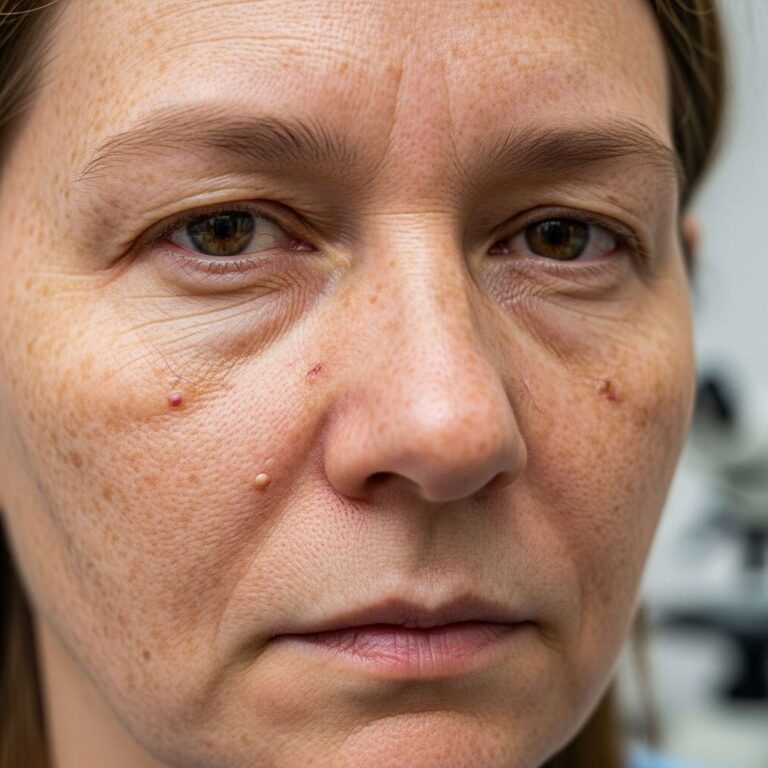
These lesions arise from the accumulation of lipid-laden macrophages, known as foam cells, in the dermis. Eruptive xanthomas typically emerge suddenly as crops of 1-4 mm dome-shaped papules with an erythematous halo, often on extensor surfaces of extremities, buttocks, and occasionally the face or trunk. They are pruritic or tender in early stages and signal significant cardiovascular risk due to associated dyslipidemias.
Pathologically, the condition reflects a secondary response to elevated chylomicrons and very low-density lipoproteins (VLDL), distinguishing it from other xanthomas like tuberous or tendinous types which relate more to hypercholesterolemia. Recognition of eruptive xanthoma prompts urgent lipid profiling, as untreated cases heighten risks of pancreatitis and atherosclerosis.
Clinical features
Eruptive xanthomas manifest acutely, often within days to weeks, as numerous small (1-5 mm) shiny papules that are yellow-orange with surrounding erythema. Lesions favor extensor arms, legs, buttocks, and hands but can generalize.
- Appearance: Dome-shaped papules, 1-4 mm, polychromatic (yellow, pink, red-brown); early halo of redness fades over weeks.
- Symptoms: Pruritus, tenderness; rarely painful unless inflamed. Koebner phenomenon reported.
- Distribution: Extensor surfaces predominant; buttocks, shoulders common; oral mucosa or face in severe cases.
- Progression: Erupt suddenly post-trigger (e.g., surgery, infection); regress in 1-2 weeks with lipid control, leaving hyperpigmentation.
Case example: A 24-year-old female developed lesions post-appendectomy, with triglycerides markedly elevated, resolving after dietary intervention.
Pathogenesis
The core pathology stems from hypertriglyceridemia, where serum triglycerides exceed 1000-2000 mg/dL, leading to chylomicronemia. Lipoprotein lipase (LPL) deficiency or dysfunction impairs triglyceride hydrolysis, causing foam cell infiltration into skin.
- Primary causes: Familial (LPL deficiency, apoC-II defects); rare, onset in childhood.
- Secondary causes: Type 2 diabetes (most common), obesity, hypothyroidism, alcohol excess, nephrotic syndrome, chronic renal failure, medications (estrogens, retinoids).
- Mechanism: Neutral fat droplets deposit in histiocytes, forming foam cells; endothelial disruption allows macrophage lipid uptake.
In diabetes-linked cases, insulin resistance exacerbates VLDL production. Genetic screening identifies familial forms, but most are acquired.
Histopathology
Microscopic examination reveals diagnostic features in the superficial to mid-dermis. Skin biopsy is confirmatory, showing:
- Foam cells: Scattered collections of lipid-laden histiocytes with vacuolated cytoplasm; Touton giant cells occasional.
- Infiltrate: Lymphocytes, neutrophils, eosinophils in early lesions; fibrosis in resolving ones.
- Epidermis: Normal or mildly hyperkeratotic; no atypia.
- Stains: Oil Red O positive for lipids; PAS highlights vacuoles.
Early lesions feature perivascular inflammation; mature ones show dense foam cell nodules. Differential includes xanthogranuloma (more Touton cells, deeper).
| Feature | Eruptive Xanthoma | Other Xanthomas |
|---|---|---|
| Foam Cells | Superficial dermis, scattered | Deeper in tuberous/tendinous |
| Inflammation | Neutrophils/eosinophils early | Minimal |
| Giant Cells | Rare Touton | Prominent in juvenile XG |
Diagnosis
Diagnosis integrates clinical eruption pattern, lipid panel (triglycerides >1000 mg/dL), and biopsy. Lipemia retinalis supports severe cases.
- Laboratory: Fasting lipids, glucose, thyroid function, renal panel.
- Imaging: Ultrasound for pancreatitis risk.
- Genetic: LPL/apoC-II assays if familial suspected.
Differentials: Xanthoma disseminatum (normolipemic, trunk), juvenile xanthogranuloma (children, solitary), eruptive histiocytoma (no lipids).
Treatment
Treatment targets underlying hyperlipidemia; lesions regress spontaneously with control.
- Lifestyle: Low-fat diet (<15% calories fat), weight loss, alcohol abstinence, exercise.
- Pharmacologic: Fibrates (fenofibrate), omega-3 fatty acids, niacin; statins if mixed dyslipidemia. Insulin for diabetes.
- Acute: Plasmapheresis for triglycerides >5000 mg/dL to avert pancreatitis.
- Lesional: Rarely needed; topical steroids for pruritus.
Monitoring: Serial lipids every 4-6 weeks; cardiovascular risk stratification essential.
Differential diagnosis
| Condition | Key Features | Lipid Levels |
|---|---|---|
| Tuberous Xanthoma | Larger firm nodules on elbows/knees | High cholesterol |
| Juvenile Xanthogranuloma | Orange nodules in children, self-resolve | Normal |
| Xanthoma Disseminatum | Red-brown papules, mucous membranes | Normal |
| Generalized Eruptive Histiocytoma | Crops on trunk, no lipids | Normal |
Frequently Asked Questions
Are eruptive xanthomas dangerous?
Not directly, but they indicate severe hypertriglyceridemia risking pancreatitis and heart disease. Prompt treatment mitigates risks.
Do eruptive xanthomas resolve on their own?
Yes, with lipid control; untreated, they persist or recur.
Can diabetes cause eruptive xanthoma?
Yes, common in uncontrolled type 1/2 diabetes due to elevated triglycerides.
Is biopsy always needed for diagnosis?
Typically clinical + lipids suffice; biopsy confirms if atypical.
What diet helps eruptive xanthoma?
Very low-fat (<20g/day), avoid sugars/alcohol; focus on fish, vegetables.
References
- Eruptive xanthomas – PMC – NIH — National Library of Medicine. 2014. https://pmc.ncbi.nlm.nih.gov/articles/PMC3907905/
- Eruptive xanthomas: Definition, symptoms, treatment, and more — Medical News Today. 2023. https://www.medicalnewstoday.com/articles/eruptive-xanthomas
- Eruptive Xanthoma – Medcom — Medcom. 2001. https://medcomhk.com/hkdvb/pdf/200112-06.pdf
- Eruptive xanthoma: Warning sign of systemic disease — Cleveland Clinic Journal of Medicine. 2015-10-01. https://www.ccjm.org/content/83/10/715
- Xanthomas Treatment Palmdale Dermatologist — Palmdale Dermatology. 2023. https://palmdaledermatology.com/medical-dermatology/xanthomas/
- Xanthoma: What It Is and How to Treat It — WebMD. 2023. https://www.webmd.com/skin-problems-and-treatments/what-is-xanthoma
- Eruptive xanthoma | MDedge – The Hospitalist — The Hospitalist. 2023. https://blogs.the-hospitalist.org/content/eruptive-xanthoma-1
Similar Articles


Read full bio of medha deb