Erysipeloid Essentials: Causes, Symptoms, And Treatment
Acute bacterial skin infection from Erysipelothrix rhusiopathiae, common in animal handlers, with self-limiting cutaneous forms.

What is erysipeloid?
Erysipeloid is an acute, usually self-limiting bacterial infection of the skin and occasionally other organs, caused by the gram-positive bacillus Erysipelothrix rhusiopathiae. This zoonotic pathogen is ubiquitous in nature, found in decaying animal matter, saltwater, freshwater environments, and commonly associated with fish, shellfish, poultry, and mammals. It primarily affects individuals with occupational exposure, such as butchers, fishmongers, slaughterhouse workers, farmers, veterinarians, and cooks, entering through minor skin abrasions or penetrating injuries.
The infection manifests most commonly as a localised cutaneous form, characterised by a distinctive violaceous, indurated plaque with a raised edge, often on the fingers or hands. Rarely, it progresses to generalised cutaneous involvement or systemic disease, including septic arthritis, endocarditis, or multisystem involvement. Despite its name suggesting similarity to erysipelas (a streptococcal infection), erysipeloid is distinct in aetiology, clinical evolution, and management.
Historically recognised since the late 19th century, erysipeloid was first described in 1884 by Danish veterinarian Daniel Cornelius Danielssen in fish handlers. Its slow progression and occupational pattern differentiate it from more acute cellulitis. Incidence is low but underreported due to self-resolution in mild cases; occupational health surveillance highlights risks in meat and seafood processing industries worldwide.
Who gets erysipeloid?
Erysipeloid predominantly affects adults engaged in occupations involving handling of raw animal products or marine life. High-risk groups include:
- Fishmongers, fishermen, and shellfish processors (most common due to E. rhusiopathiae prevalence in marine environments).
- Butchers, abattoir workers, and meat packers.
- Poultry and swine farmers or processors.
- Veterinarians and animal handlers.
- Cooks, grocers, and food preparers exposed to contaminated products.
Men are affected more frequently than women, reflecting occupational disparities. Children and non-exposed individuals rarely develop it, except via rare person-to-person transmission or contaminated wounds. Immunocompromised patients (e.g., diabetes, alcoholism, asplenia) face higher risks of dissemination.
Geographically, cases cluster in coastal regions and areas with intensive animal husbandry. Outbreaks have been documented in seafood processing plants, with attack rates up to 20% among injured workers without prophylaxis.
Causes and pathophysiology
Erysipelothrix rhusiopathiae is a thin, encapsulated, non-motile, non-spore-forming, facultatively anaerobic gram-positive rod. It exists in three serological variants (1a, 1b, 2), with serovar 1b most virulent in humans. As a saprophyte, it thrives on decaying organic matter but acts as an opportunistic pathogen in mammals, birds, reptiles, and fish.
Infection occurs via percutaneous inoculation through cuts, punctures, or abrasions contaminated with infected tissues. The organism evades phagocytosis via its capsule and virulence factors like surface protective antigen A (SpaA), neuraminidase, hyaluronidase, and cell wall proteins, enabling intracellular survival in macrophages. This allows local proliferation and rare haematogenous dissemination.
Once in skin, bacteria induce a localised inflammatory response without pus formation, leading to the characteristic cellulitis. Bacteraemia is infrequent (less than 5%) but can seed distant sites like joints (septic arthritis in 50% of systemic cases) or heart valves (endocarditis, often right-sided).
Clinical features
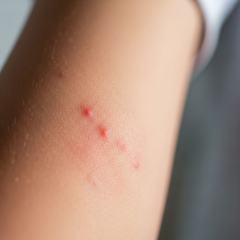
Symptoms emerge 2–7 days post-inoculation. The localised form (90–95% of cases) presents as:
- Well-demarcated, raised, violaceous or purple-red plaque with sharp advancing edge and central clearing.
- Intense pruritus, burning, or throbbing pain; minimal systemic symptoms.
- Affected site typically finger webspaces, backs of hands, wrists; swelling impairs function.
- Duration 2–4 weeks if untreated; spontaneous resolution common.
- Lymphangitis or regional lymphadenopathy in ~30%.
Generalised cutaneous form (rare): Multiple spreading lesions, vesicles, or bullae; mild systemic signs.
Systemic erysipeloid (very rare): Fever, chills, arthralgias, myalgias; complications include:
- Septic arthritis (knees, shoulders).
- Infective endocarditis (vancomycin-resistant, poor prognosis if delayed).
- Rarely: meningitis, pneumonia, osteomyelitis.
Diagnosis
Clinical suspicion in endemic occupations with compatible lesions suffices for presumptive diagnosis. Confirmation requires:
- Culture: Full-thickness skin biopsy, lesion swab, or aspirate on blood agar (growth in 48–72 hours; characteristic ‘pinpoint’ colonies).
- PCR: For rapid detection in culture-negative cases.
- Histology: Intraepidermal microabscesses, dermal oedema, sparse neutrophils; Gram-positive rods.
- Blood cultures if systemic symptoms.
- Differential: Cellulitis (streptococcal), sporotrichosis, contact dermatitis, herpes zoster, necrotising fasciitis.
| Condition | Distinguishing Features |
|---|---|
| Erysipelas | More acute, streptococcal, facial predilection, responds to penicillin. |
| Cellulitis | Diffuse, tender, purulent; fever common early. |
| Sporotrichosis | Lymphocutaneous nodules along lymphatics; fungal. |
| Contact dermatitis | Itchy, no induration, history of allergen exposure. |
Treatment
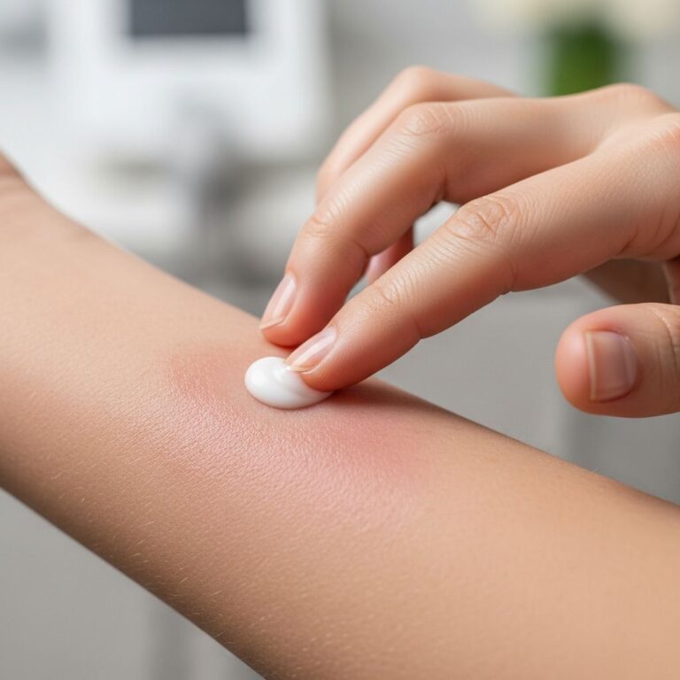
Localised cutaneous erysipeloid often resolves without intervention in 3–4 weeks, but antibiotics hasten recovery and prevent spread. First-line:
- Penicillin V/G 500mg QID or IM procaine penicillin (7–14 days).
- Cephalosporins (e.g., cephalexin) for penicillin-allergic.
- Alternatives: Ciprofloxacin, tetracyclines, erythromycin + rifampicin.
Systemic cases require IV penicillin G (4 weeks minimum); endocarditis needs 6 weeks IV + surgery if valve destruction. E. rhusiopathiae resists vancomycin, critical for empiric endocarditis therapy.
Supportive: Elevation, analgesics, wound care. Monitor for dissemination in at-risk patients.
What is the prognosis for erysipeloid?
Excellent for cutaneous forms (>95% resolve fully without scarring). Untreated localised cases heal in 2–4 weeks. Antibiotics reduce duration to 1 week. Systemic complications carry 10–50% mortality if endocarditis develops, especially pre-antibiotic era. Recurrence rare unless re-exposure.
Prevention of erysipeloid
Occupational prevention key:
- Wear puncture-resistant gloves, aprons when handling raw products.
- Immediate wound cleansing with soap/water, povidone-iodine; prophylactic antibiotics (penicillin V 500mg BID x 3 days) for high-risk injuries.
- Education in slaughterhouses/seafood plants.
- Vaccination under research (SpaA-based).
Frequently asked questions
What does erysipeloid look like?
A sharply demarcated, raised, purple-red plaque with burning pain and itching, often on hands.
Is erysipeloid contagious?
No, not person-to-person; requires direct inoculation from contaminated sources.
How long does erysipeloid last?
Untreated: 2–4 weeks; treated: 5–7 days.
Does erysipeloid need antibiotics?
Recommended to speed healing and prevent spread, though self-limiting.
Can erysipeloid cause permanent damage?
Rarely; mainly in untreated endocarditis or arthritis.
References
- Erysipeloid – Infectious Diseases — Merck Manual Professional Edition. 2023. https://www.merckmanuals.com/professional/infectious-diseases/gram-positive-bacilli/erysipeloid
- Erysipeloid – Infectious Diseases — MSD Manual Professional Edition. 2023. https://www.msdmanuals.com/professional/infectious-diseases/gram-positive-bacilli/erysipeloid
- Erysipeloid — MedlinePlus Medical Encyclopedia. 2024-01-15. https://medlineplus.gov/ency/article/000632.htm
- Erysipeloid — DermNet NZ. 2023. https://dermnetnz.org/topics/erysipeloid
- Erysipeloid: a review — Clinical and Experimental Dermatology, Oxford Academic. 2009-10-20. https://academic.oup.com/ced/article/34/8/859/6625401
- ERYSIPELOID: REPORT OF A CASE — JAMA Dermatology. 1950. https://jamanetwork.com/journals/jamadermatology/fullarticle/519509
Similar Articles


Read full bio of Sneha Tete