Erythema Dyschromicum Perstans: Diagnosis and Management
Understanding ashy dermatosis: Clinical features, diagnosis, and treatment options for this persistent skin condition.

Erythema Dyschromicum Perstans: Clinical Overview
Erythema dyschromicum perstans (EDP), commonly referred to as ashy dermatosis, is a form of acquired dermal macular hyperpigmentation characterized by well-circumscribed round to oval or irregular patches on the skin. This chronic dermatological condition predominantly affects individuals with intermediate to dark skin types and exhibits a notable female predominance. The disease is characterized by persistent, gray-blue or ashy-brown macules that typically appear on the trunk, face, neck, arms, and legs, though the distribution can vary considerably among affected individuals.
The condition is notable for its chronic nature and resistance to conventional therapeutic interventions. Patients frequently experience lesions that may persist unchanged for years, although some cases eventually resolve spontaneously. The asymptomatic nature of EDP in most cases, combined with its cosmetically significant appearance, makes it a source of considerable concern for affected patients despite the absence of physical symptoms such as pruritus or pain.
Clinical Presentation and Characteristics
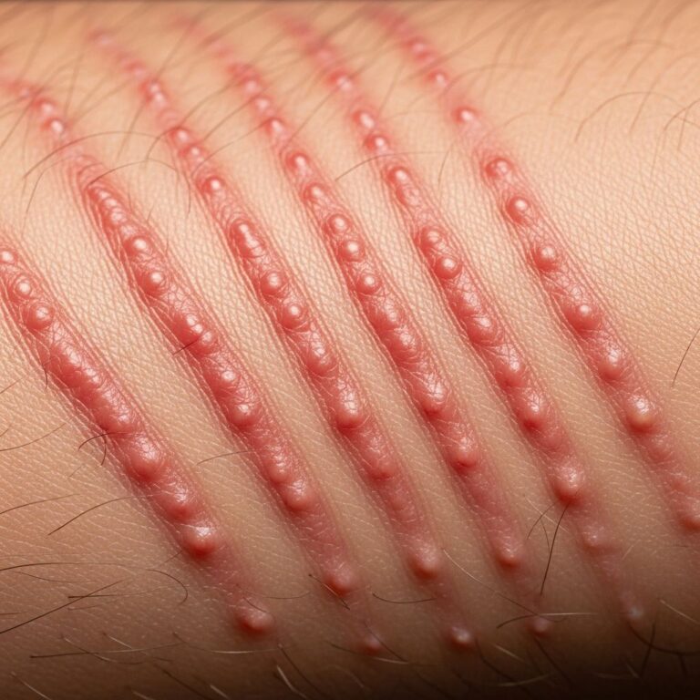
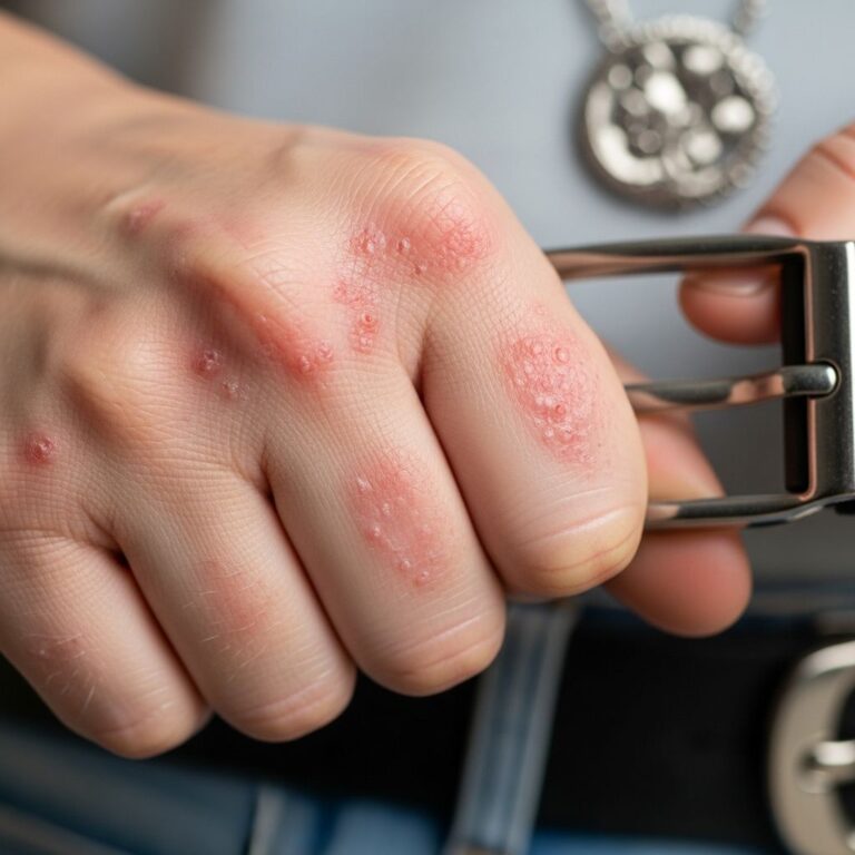
Patients with erythema dyschromicum perstans typically present with subacute to chronic, asymptomatic lesions that are symmetric in distribution. The characteristic gray-blue or ashy-brown coloration distinguishes EDP from other hyperpigmentary disorders. These macules may appear with an erythematous halo or active red border in some cases, particularly at the periphery of lesions where active inflammation is present.
The morphology and distribution of EDP lesions include:
- Well-demarcated, round to oval patches or irregular plaques
- Gray-blue to ashy-brown coloration with variable intensity
- Potential erythematous borders indicating active lesions
- Predominant truncal involvement with extension to face, neck, extremities, and legs
- Potential for widespread distribution across multiple body sites
- Typically symmetric presentation patterns
The active border of lesions often represents the inflammatory phase, while the central portions display established hyperpigmentation from dermal melanophages and pigment incontinence. Most patients report the absence of symptoms, and classic pruritis associated with other inflammatory conditions like lichen planus is typically absent.
Etiology and Pathophysiology
The precise etiology of erythema dyschromicum perstans remains unclear, though research has identified several non-conclusive associations that may trigger or contribute to disease development. Proposed etiological factors include endocrinopathies, exposure to chemical substances, association with vitiligo, and chronic hepatitis C infection. Recent case reports have documented associations with COVID-19 vaccination, expanding the potential trigger list.
The pathophysiology of EDP involves a complex interplay between inflammatory and pigmentary changes. The condition appears to result from a lichenoid inflammatory response that disrupts normal melanin processing and causes abnormal deposition of melanin in dermal macrophages, leading to the characteristic gray-blue discoloration. This mechanism distinguishes EDP from other hyperpigmentary conditions and suggests a unique pathophysiological process.
Histopathological Features
Histological examination reveals distinct features that aid in diagnosis, though findings must be interpreted alongside clinical presentation. The histology of EDP can be subdivided into active (early) and inactive (late) lesions, each demonstrating characteristic microscopic findings.
Active lesions typically demonstrate:
- Basal vacuolar degeneration at the dermal-epidermal junction
- Edematous papillary dermis with pronounced lymphocytic infiltration
- Perivascular mononuclear cell infiltrates
- Increased epidermal melanin content
- Minor vacuolar degeneration of the basal layer
Inactive or established lesions characteristically show:
- Melanophages and pigment incontinence in the dermis without active inflammation
- Post-inflammatory changes with melanin-filled macrophages responsible for distinctive gray-blue coloration
- Dermal melanophages and hemosiderin deposits in isolation
- Increased melanin within the epidermis causing the characteristic discoloration
A significant percentage of cases reveal melanophages, lymphocytic infiltrates, and basal vacuolar degeneration, with notable histologic overlap observed with lichen planus pigmentosus (LPP). However, a diagnosis of EDP should not be based on histologic findings alone; clinical history, morphology, and distribution are essential for accurate differentiation from LPP.
Diagnostic Approach
Diagnosis of erythema dyschromicum perstans relies on a combination of clinical assessment and supportive diagnostic findings. In some cases, the clinical picture may be sufficiently characteristic to establish diagnosis without additional testing. However, when diagnosis is uncertain, skin biopsy provides valuable confirmatory information.
A punch biopsy taken from the border of pigmentation reveals characteristic findings including superficial perivascular lymphocytic infiltrate with vacuolar interface change in the papillary dermis consistent with EDP. The biopsy should ideally sample the active peripheral border where inflammatory changes are most prominent, as central portions may show only post-inflammatory features.
Differential diagnosis is crucial, particularly distinguishing EDP from:
- Lichen planus pigmentosus (LPP) – where sun-avoidance is recommended and UVB exposure typically worsens disease
- Other acquired hyperpigmentary disorders
- Post-inflammatory hyperpigmentation from preceding inflammatory conditions
Laboratory studies, including complete blood count, iron studies, and hemoglobin A1C levels, are typically unremarkable and serve primarily to exclude other systemic conditions.
Treatment Options and Clinical Efficacy
Erythema dyschromicum perstans is notably resistant to conventional treatments, and no consistently effective single therapy currently exists. Treatment decisions should be individualized based on patient tolerance, disease extent, and response to initial interventions. The therapeutic approach emphasizes matching treatment choice to potential side effect profiles.
Topical Treatments
Topical corticosteroids represent first-line therapy for localized disease. These agents are applied directly to affected areas to reduce inflammation and pruritus, though efficacy in resolving established lesions varies considerably. Topical corticosteroid responses are often unsatisfactory when used as monotherapy.
Tacrolimus, a macrolide antibiotic with immunosuppressive properties, has emerged as the most promising topical treatment option. This medication exerts its effects principally through inhibition of calcium-dependent events, including interleukin 2 gene transcription, nitric oxide synthase activation, cell degranulation, and apoptosis. Studies demonstrate that tacrolimus produces effective therapeutic results with minimal side effects, making it an attractive option for patients seeking treatment.
Ruxolitinib, a novel topical treatment representing a Janus kinase (JAK) inhibitor, has shown promise in recent case reports. A 57-year-old patient treated with topical ruxolitinib 1.5% cream twice daily demonstrated improvement of lesions after one month of use, suggesting potential efficacy for this emerging therapy.
Systemic Treatments
Clofazimine has been identified as the most successful systemic treatment option. This antimicrobial agent has demonstrated effectiveness in multiple case reports; however, its use is frequently limited by significant to intolerable side effects that compromise patient tolerance.
Other oral medications that have been used with variable success include:
- Dapsone – improved lesions in some patients but lesions frequently recurred after discontinuation
- Griseofulvin – provided unsatisfactory results with recurrence after treatment cessation
- Hydroxychloroquine – successful in limited case reports
- Isoniazid – demonstrated benefit in select cases
- Antimalarial agents – reported use without clear evidence of consistent benefit
- Corticosteroids – variable efficacy in small patient populations
- Isotretinoin – demonstrated unsatisfactory results with recurrence after discontinuation
- Minocycline – helpful in some patients, though efficacy remains inconsistent
Phototherapy
Narrowband UVB (NB-UVB) therapy represents a promising treatment approach that distinguishes EDP from LPP in terms of therapeutic response. Unlike LPP where sunlight exposure and UVB therapy exacerbate disease, UVB therapy—either through natural sunlight or controlled NB-UVB exposure—improves EDP in reported cases.
This differential response to UVB therapy suggests that EDP and LPP represent separate entities with distinct pathophysiology. In one notable case, a patient initiated on NB-UVB therapy at 300 mJ three times weekly, with 10-15% incremental increases per session, experienced resolution of erythema and pruritus with significant hyperpigmentation decrease after two months. Extended follow-up demonstrated sustained resolution at four years, marking the first reported case of prolonged EDP remission following NB-UVB therapy.
Laser therapy, conversely, has proven largely ineffective and may cause postinflammatory hyperpigmentation and fibrosis, making it a poor treatment choice for this population.
Treatment Comparison Table
| Treatment | Type | Efficacy | Side Effects |
|---|---|---|---|
| Tacrolimus | Topical | Promising | Minimal |
| NB-UVB | Phototherapy | Promising | Minimal |
| Clofazimine | Systemic | Most Effective | Significant-to-Intolerable |
| Dapsone | Systemic | Variable | Moderate |
| Griseofulvin | Systemic | Unsatisfactory | Moderate |
| Topical Corticosteroids | Topical | Variable | Minimal-Moderate |
| Laser | Procedural | Ineffective | PIH, Fibrosis |
Prognosis and Long-Term Outcomes
The prognosis of erythema dyschromicum perstans is variable and unpredictable. The condition is characterized by persistent lesions that may remain stable for extended periods, ranging from months to years. In some instances, lesions eventually clear spontaneously without intervention, though this represents the minority of cases.
The recalcitrant nature of EDP, combined with its cosmetically significant appearance and variable response to treatment, necessitates realistic patient counseling regarding disease trajectory and therapeutic expectations. Treatment responses, when they occur, may require extended duration before clinical improvement becomes apparent, necessitating patient commitment to prolonged therapeutic courses.
Frequently Asked Questions
Q: What is the difference between erythema dyschromicum perstans and lichen planus pigmentosus?
A: While both conditions can show histologic overlap with lichenoid infiltrates, they differ significantly in their response to sun exposure. EDP improves with UVB therapy, whereas LPP typically worsens with sun exposure, making sun-avoidance essential for LPP management. Additionally, clinical history, morphology, and distribution patterns distinguish these entities.
Q: Is erythema dyschromicum perstans itchy or painful?
A: No, EDP is characteristically asymptomatic. Most patients do not experience pruritus, pain, or other subjective symptoms, distinguishing it from inflammatory conditions like classic lichen planus.
Q: Can erythema dyschromicum perstans be cured?
A: There is no consistently effective cure for EDP. While some cases eventually resolve spontaneously, others persist for years. Current treatments aim to improve appearance rather than provide definitive cure, with NB-UVB and topical tacrolimus showing the most promising results with minimal side effects.
Q: Who is most likely to develop erythema dyschromicum perstans?
A: EDP predominantly affects individuals with intermediate to dark skin types and exhibits a notable female predominance. The condition can develop at various ages but appears more common in specific ethnic populations.
Q: What should I do if my lesions don’t improve with treatment?
A: If initial treatments prove ineffective, consultation with a dermatologist is essential to explore alternative options. Treatment-resistant cases may benefit from combination approaches or newer therapies such as topical ruxolitinib, though larger clinical studies are needed to confirm efficacy.
References
- Erythema dyschromicum perstans: A case report and systematic review of literature — National Center for Biotechnology Information (NCBI/PMC). 2018. https://pmc.ncbi.nlm.nih.gov/articles/PMC6322153/
- Successful Management of Erythema Dyschromicum Perstans Following Topical Ruxolitinib Therapy — Journal of Drugs in Dermatology. 2023. https://jddonline.com/articles/successful-management-of-erythema-dyschromicum-perstans-following-topical-ruxolitinib-therapy-S1545961623P0297X
- Erythema Dyschromicum Perstans vs Lichen Planus — Perri Dermatology. https://perridermatology.com/dr-perris-blog/lichen-planus-erythema-dyschromicum-perstans/
- Erythema dyschromicum perstans (ashy dermatosis) — DermNet New Zealand. https://dermnetnz.org/topics/erythema-dyschromicum-perstans
- Erythema Dyschromicum Perstans — Consultant360. https://www.consultant360.com/articles/erythema-dyschromicum-perstans
Similar Articles


Read full bio of Sneha Tete