Erythrasma: Diagnosis, Treatment, Prevention Expert Guide
Comprehensive guide to erythrasma: causes, symptoms, diagnosis, treatment, and prevention of this common bacterial skin infection.

What is erythrasma?
Erythrasma is a superficial chronic bacterial infection of the skin caused by the gram-positive bacillus Corynebacterium minutissimum. It typically manifests as well-defined pink or brown patches with fine scaling in intertriginous areas—regions where skin surfaces rub together, such as the armpits, groin, and interdigital spaces between toes. This condition is often asymptomatic but can cause mild itching or burning, particularly in the groin. Erythrasma is more prevalent in warm, humid climates and among adults, especially males in groin areas and females between toes.
Unlike fungal infections, erythrasma results from overgrowth of normal skin flora in moist environments. It is classified into three main types: interdigital (toe webs), intertriginous (skin folds), and generalized or disciform (widespread on trunk and limbs). The infection thrives in occluded, sweaty areas, leading to maceration and fissuring. On darker skin tones, patches may appear hypopigmented with hyperpigmented borders.
Who gets erythrasma?
Erythrasma affects individuals across demographics but shows distinct patterns. It is more common in adults than children, with a higher incidence in males for groin involvement and females for interdigital areas. Risk factors include:
- Living in tropical or humid climates, where moisture promotes bacterial proliferation.
- Obesity, which creates more skin folds prone to friction and occlusion.
- Diabetes mellitus, particularly type 2, impairing skin immunity and increasing susceptibility; widespread forms may signal undiagnosed diabetes.
- Excessive sweating (hyperhidrosis) and poor hygiene.
- Immunocompromised states, advanced age, or skin of color.
Prevalence is higher in overweight individuals and those with chronic conditions favoring moist skin environments. It is rarely seen in dry climates or young children.
Causes
The causative agent is Corynebacterium minutissimum, a normal commensal skin bacterium that becomes pathogenic in predisposing conditions. This gram-positive, non-spore-forming, aerobic or facultative anaerobic bacillus produces porphyrins, responsible for the characteristic coral-red fluorescence under Wood’s lamp. Overgrowth occurs in warm, moist, occluded areas due to:
- High humidity and temperature disrupting skin barrier.
- Friction in skin folds macerating the stratum corneum.
- Underlying conditions like diabetes altering local immunity.
Historical note: First described in 1859, initially misattributed to fungi; C. minutissimum identified later. It may coexist with fungal infections like tinea or Candida, complicating diagnosis.
Clinical features
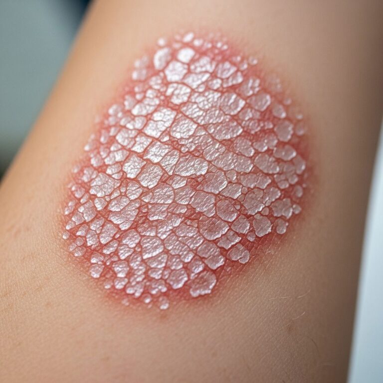
Erythrasma presents with sharply demarcated patches of pink to brown discoloration, fine scaling, superficial fissures, and macerated, wrinkled skin resembling ‘cigarette paper’. Patches expand slowly without significant inflammation. Common sites include:
- Interdigital: Between toes (most common foot infection), causing cracking and scaling; often coexists with fungal toe web infection.
- Intertriginous: Groin, axillae, inframammary folds, intergluteal cleft, umbilicus, abdominal creases; prevalent in diabetics.
- Generalized/disciform: Rare, plaque-like on trunk/limbs; more in tropical-dwelling women, diabetes-associated.
Symptoms are often absent, but mild pruritus, burning, or odor may occur, especially groin. On darker skin: hypopigmented center with darker edges. Images typically show uniform patches without pustules or vesicles.
Diagnosis
Diagnosis relies on clinical appearance, confirmed by:
- Wood’s lamp examination: Bright coral-red fluorescence due to bacterial porphyrins (pathognomonic).
- Clinical inspection: Uniform patches in folds without satellite lesions (vs. candida).
- Microscopy: Skin scrapings in 20% KOH show gram-positive filaments; no hyphae.
- Culture: Rarely needed; grows C. minutissimum on blood agar.
- Biopsy: Confirms superficial dermal bacterial colonization if atypical.
Differential includes tinea, candida intertrigo, psoriasis, contact dermatitis, seborrheic dermatitis. Wood’s lamp distinguishes: no fluorescence in fungi, yellow-green in pseudomonas.
Treatment
Most cases respond to topical therapy; extensive disease requires systemic antibiotics.
Topical treatments
- Antiseptics: Clioquinol-hydrocortisone, Whitfield ointment (benzoic/salicylic acids).
- Topical antibiotics: Mupirocin, clindamycin, erythromycin, fusidic acid; apply BID for 2 weeks.
Systemic antibiotics
For widespread or recalcitrant cases:
- Erythromycin 250mg QID or tetracycline 250mg QID for 2 weeks.
- Alternatives: clarithromycin, doxycycline.
Adjuncts: Keep areas dry (talcum powder), loose clothing. Photodynamic therapy (red light 635nm) for resistant cases. Recurrence common without addressing predispositions.
Complications
Usually self-limiting, but potential issues include:
- Secondary contact dermatitis, lichenification, postinflammatory hyperpigmentation.
- Coinfections with dermatophytes, yeasts, other bacteria.
- Rare systemic: abscess, cellulitis, endocarditis, pyelonephritis in immunocompromised.
No scarring typically; prompt treatment prevents spread.
Prevention
Focus on reducing moisture and friction:
- Maintain hygiene: daily washing, thorough drying of folds.
- Wear breathable fabrics, loose clothing.
- Absorbent powders (non-cornstarch) in folds.
- Weight management, diabetes control.
- Avoid occlusive ointments.
Prophylactic topical antibiotics for recurrences.
Frequently asked questions
Is erythrasma contagious?
Erythrasma is not typically contagious person-to-person but spreads via autoinoculation in predisposed individuals.
Does erythrasma go away on its own?
It is often self-limiting but persists chronically without treatment; addressing moisture resolves many cases.
How long does treatment take?
Topical therapy clears patches in 1-2 weeks; systemic for extensive disease.
Can erythrasma affect children?
Rare in children; primarily adults.
Is Wood’s lamp always needed for diagnosis?
Helpful but not mandatory; clinical features suffice often.
| Type | Common Sites | Features | Risk Groups |
|---|---|---|---|
| Interdigital | Toe webs | Maceration, fissuring | Females, foot sweat |
| Intertriginous | Groin, axillae, folds | Scaly pink-brown patches | Males, diabetics, obese |
| Disciform | Trunk, limbs | Scaly plaques, Wood’s glow | Tropical women, diabetes |
References
- Erythrasma: Causes, Symptoms, and Treatment — WebMD. 2023. https://www.webmd.com/skin-problems-and-treatments/what-is-erythrasma
- Erythrasma — DermNet NZ. 2024-01-15. https://dermnetnz.org/topics/erythrasma
- Erythrasma — ADAM Health Encyclopedia (sbrmc.adam.com). 2023. https://sbrmc.adam.com/content.aspx?productid=117&pid=1&gid=001470
- Erythrasma — Skin Solutions Dermatology. 2024. https://www.skinsolutionsderm.com/our-services/medical-dermatology/rash/erythrasma/
- Erythrasma – StatPearls — NCBI Bookshelf. 2023-08-07. https://www.ncbi.nlm.nih.gov/books/NBK513352/
Similar Articles


Read full bio of Sneha Tete