Erythropoietic Protoporphyria Pathology: Clinical Insights
Detailed pathology of erythropoietic protoporphyria, a rare photodermatosis causing painful skin reactions to sunlight exposure.

Erythropoietic protoporphyria (EPP) is a rare inherited photodermatosis characterized by partial blockage of the final step in haem biosynthesis, leading to accumulation of protoporphyrin IX (PPIX) predominantly in red blood cells, plasma, liver, and skin. This accumulation causes acute painful photosensitivity upon visible light exposure without preceding erythema or vesicles, distinguishing it from other porphyrias. Pathologically, EPP manifests with unique histopathological changes in acutely and chronically photoexposed skin, reflecting photosensitizer-induced damage.
Histopathology
The histopathology of EPP varies depending on whether the skin biopsy is taken from an acute reaction site or areas of chronic photodamage. In acute lesions, which occur minutes to hours after visible light exposure, epidermal changes are minimal. There is subtle epidermal cell degeneration without significant lymphocytic infiltrate or vascular damage initially. Parakeratosis and mild spongiosis may be observed, but the hallmark is the presence of hyaline eosinophilic material around superficial dermal vessels, representing laminated periodic acid-Schiff (PAS)-positive material and basement membrane thickening.
Electron microscopy reveals more specific features in acute reactions, including endothelial cell degeneration with cytoplasmic vacuolation and swelling of perivascular cell processes. Protoporphyrin deposits appear as electron-dense, laminated bodies within lysosomes of endothelial cells, pericytes, and Schwann cells. These findings correlate with the rapid onset of pain due to photodynamic activation of PPIX in vessel walls.
In chronic photoexposed skin, particularly on the dorsum of hands and face, the epidermis shows hyperkeratosis, acanthosis, and elongation of rete ridges. The papillary dermis exhibits increased elastic tissue with thick, clumped fibres (solar elastosis) and a marked increase in PAS-positive material around vessels. Fibrosis replaces normal dermal structures, and eccrine ducts may show dilated lumina with PAS-positive walls. These changes result from repeated phototoxic injury over years.
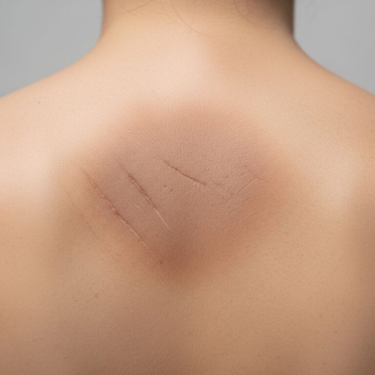
Histopathology images
- Low power view: Acute EPP lesion demonstrating subtle epidermal changes and prominent perivascular hyaline material in the superficial dermis.
- High power view: Chronic photodamaged skin showing epidermal acanthosis, papillary dermal fibrosis, and thickened vessel walls with PAS-positive deposits.
- Electron micrograph: Laminated electron-dense protoporphyrin inclusions in endothelial cell lysosomes from acutely exposed skin.
These images illustrate the progression from acute vascular damage to chronic fibrotic changes characteristic of EPP photodamage.
Pathogenesis
EPP results from compound heterozygous mutations in the ferrochelatase gene (FECH) on chromosome 18q21, which catalyses the insertion of ferrous iron into PPIX to form heme. Typically, patients inherit one severe loss-of-function mutation and one hypomorphic (low-expression) allele, reducing enzyme activity to 15-25% of normal. Rarely, gain-of-function mutations in erythroid ALA synthase 2 (ALAS2) cause X-linked protoporphyria (XLP) with similar manifestations.
Reduced ferrochelatase activity leads to massive accumulation of free PPIX in bone marrow reticulocytes, erythrocytes, plasma, bile, and skin. PPIX strongly absorbs visible light at 410nm (Soret band) and emits fluorescence at 634nm, serving as a diagnostic marker. Upon light exposure, PPIX generates singlet oxygen and other reactive oxygen species via photodynamic reaction, primarily damaging vascular endothelium in the superficial dermis.
The pain arises from C-nociceptor activation by phototoxins, hydrogen peroxide, and bradykinin release, without initial inflammation. Chronic exposure leads to dermal fibrosis as repeated endothelial injury triggers collagen deposition. Approximately 5% of patients develop protoporphyrin-induced cholestatic liver disease progressing to cirrhosis in 2-5%, with higher erythrocyte PPIX levels predicting risk.
Clinical features
Symptoms typically begin in infancy or early childhood, with children crying after brief sunlight exposure. Patients experience prodromal tingling, burning, or itching within minutes, progressing to severe pain lasting 24-96 hours if exposure continues. Notably, objective signs like erythema and edema are often absent or minimal, leading to frequent misdiagnosis as psychosomatic.
Chronically exposed sites develop waxy scars, skin thickening, and pitted scars on the dorsum of hands and over knuckles. Facial skin shows leathery texture and cheilitis with shallow radial scars around the lips. Palms and soles may develop hyperkeratotic lesions mimicking pachydermoperiostosis. Gallstones occur in 20-30% due to PPIX precipitation in bile.
| Clinical Feature | Pathological Finding | Mechanism |
|---|---|---|
| Acute pain without erythema | Vascular endothelial damage | PPIX photodynamic reaction |
| Knuckle scarring | Papillary dermal fibrosis | Repeated phototoxic injury |
| Cheilitis | Epidermal acanthosis, PAS+ vessels | Chronic lip exposure |
| Gallstones | Biliary PPIX crystals | Protoporphyrin insolubility |
Diagnosis
Clinical suspicion arises from childhood-onset photosensitivity without blisters or hypertrichosis. Definitive diagnosis requires biochemical confirmation: markedly elevated total erythrocyte protoporphyrin (>80μmol/L, normal <1.5) with >80% metal-free PPIX (vs zinc protoporphyrin in iron deficiency). Plasma shows characteristic fluorescence peak at 634nm.
Genetic testing identifies FECH mutations (autosomal recessive) or ALAS2 mutations (X-linked). Skin biopsy, while not diagnostic alone, shows characteristic vascular PAS-positive material supporting the diagnosis. Liver biopsy in suspected cases reveals protoporphyrin crystals and cholestasis. Blood for porphyrin analysis must be protected from light.
Differential diagnosis
- Other porphyrias: Variegate porphyria and hereditary coproporphyria cause blistering, not acute pain.
- Polymorphic light eruption: Develops after hours, shows vesicles and lymphocytic infiltrate.
- Hydroa vacciniforme: Vesicles and umbilicated scars, begins later in childhood.
- Solar urticaria: Immediate wheals, responds to antihistamines.
- Kindler syndrome: Blisters, poikiloderma, mucosal involvement.
Disease severity classification
| Severity | Sun Tolerance | Skin Changes | Liver Risk |
|---|---|---|---|
| Mild | >1 hour | Minimal scarring | Low (<20μmol/L plasma PPIX) |
| Moderate | 15-60 min | Moderate scarring | Intermediate |
| Severe | <15 min | Marked scarring, cheilitis | High (>100μmol/L plasma PPIX) |
Frequently Asked Questions
What causes the pain in EPP?
The severe pain results from photodynamic activation of protoporphyrin in vascular endothelium, generating reactive oxygen species that stimulate C-nociceptors without initial inflammation.
Does EPP cause skin cancer?
No increased skin cancer risk despite chronic damage; the short-lived singlet oxygen primarily causes fibrosis rather than mutagenesis.
Can liver disease be prevented?
Regular monitoring of liver function and erythrocyte PPIX levels allows early intervention. Phlebotomy and ursodeoxycholic acid reduce hepatic protoporphyrin accumulation.
Is genetic testing necessary?
Recommended for confirmation, family screening, and reproductive counseling, identifying carriers with low-expression alleles.
What light sources trigger symptoms?
Visible light (400-700nm), including sunlight, fluorescent bulbs, and LED screens. UVA/UVB protection insufficient alone.
Management
Avoidance of visible light using protective clothing and window films is primary. Afamelanotide (SCENESSE®) increases eumelanin, extending light tolerance by 4-6 hours. Iron supplementation paradoxically reduces PPIX by favoring ferrochelatase activity. For liver disease, ursodeoxycholic acid promotes biliary PPIX excretion; liver transplant curative for end-stage disease.
Experimental therapies include luciferase-luciferin system to catabolize PPIX and gene therapy targeting bone marrow.
References
- Erythropoietic Protoporphyria — DermNet NZ. 2023. https://dermnetnz.org/topics/erythropoietic-protoporphyria
- Erythropoietic Protoporphyria and X-Linked Porphyria — American Porphyria Foundation. 2024-01-15. https://www.porphyria.org/epp-xlp
- Porphyria – Symptoms and causes — Mayo Clinic. 2025-06-12. https://www.mayoclinic.org/diseases-conditions/porphyria/symptoms-causes/syc-20356066
- Erythropoietic Protoporphyria and X-linked Protoporphyria — MSD Manual Professional Edition. 2025-03-01. https://www.msdmanuals.com/professional/endocrine-and-metabolic-disorders/the-porphyrias/erythropoietic-protoporphyria-and-x-linked-protoporphyria
- Erythropoietic Protoporphyria — StatPearls, NCBI Bookshelf. 2024-07-20. https://www.ncbi.nlm.nih.gov/books/NBK563141/
Similar Articles


Read full bio of Sneha Tete