Eyes: How They Work, Anatomy & Common Conditions
Understanding your eyes: Complete guide to eye anatomy, vision function, and common eye conditions.

Your eyes are remarkable light-gathering sensory organs positioned at the front of your head. They serve as the gateway to your visual world, sending signals that your brain processes to create the picture you see. Understanding how your eyes function, their complex anatomy, and the conditions that can affect them is essential for maintaining healthy vision throughout your life.
How Your Eyes Work: The Vision Process
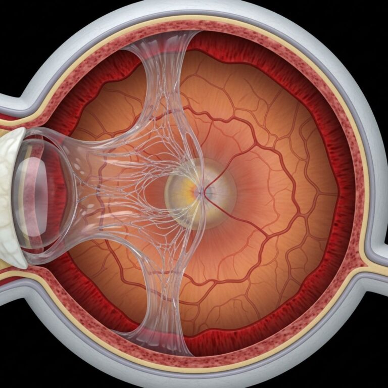
The process of seeing begins with light from the outside world. Your eye structure is perfectly designed to let light enter and pass through a series of clear components and sections, including the cornea, aqueous humor, lens, and vitreous humor. These structures work together to bend and focus light, carefully adjusting how far the light beams travel before they come into focus on the retina.
For clear vision, this focus must be precise. If the focus point is off even slightly, what you’re looking at appears blurry. Your eye has sophisticated muscles that make subtle changes to the shape of your eye, moving the focus point so it lands correctly on the retina. This automatic adjustment happens constantly throughout your day, allowing you to see clearly whether you’re looking at something close by or far away.
The Anatomy of Your Eye: Understanding Each Component
The Cornea: Your Eye’s First Lens
The cornea is the transparent, dome-shaped front part of your eyeball that covers the iris, pupil, and anterior chamber. It acts as your eye’s outermost lens, bending incoming light and directing it through the pupil toward the lens. The cornea is responsible for roughly 70 percent of your eye’s focusing power, making it one of the most critical refracting surfaces in your visual system. Remarkably, the cornea contains no blood vessels, which allows it to remain completely transparent. However, this also makes it highly susceptible to discomfort and pain when irritated.
The Sclera: The White of Your Eye
The sclera is the white part of your eye that you can see in the mirror. This tough, fibrous tissue wraps around your entire eyeball, providing structural support and maintaining your eye’s shape. The sclera serves as an attachment point for the external eye muscles that control eye movement and acts as a protective covering for the delicate internal structures of your eye.
The Conjunctiva: Protective Membrane
The conjunctiva is a thin, transparent membrane that covers the inner surface of your eyelids and extends across the white part of your eye. This protective layer works in conjunction with your tear glands to produce tears that keep your eye moist, clean, and free from infection and debris. The conjunctiva contains blood vessels and nerve endings that help protect your eye from damage.
The Iris and Pupil: Controlling Light Entry
The iris is the colored part of your eye that contains muscles controlling the pupil, which is the dark opening in the center of your iris. These structures work together to regulate the amount of light entering your eye. When you’re exposed to bright light, the iris automatically constricts the pupil to reduce light entry and protect your retina. Conversely, when lighting is dim, the iris dilates the pupil to allow more light into your eye for improved visibility.
The Aqueous Humor: Nourishing Your Eye
The aqueous humor is a clear, watery fluid that fills the front part of your eye. This fluid serves two critical functions: it nourishes the cornea and lens, which don’t have blood vessels, and it maintains the shape and pressure of your eye to keep it properly inflated. Your eye continuously produces a small amount of aqueous humor while an equal amount flows through the trabecular meshwork at the drainage angle, maintaining a balanced internal pressure.
The Lens: Fine-Tuning Your Focus
Your eye lens is a flexible, clear structure situated just behind your pupil, between the pupil and retina. Made up of clear crystalline proteins, the lens is your eye’s chance to fine-tune your focus after light has already been bent by the cornea. The lens provides approximately 30 percent of your eye’s total focusing power, while the cornea provides the remaining 70 percent.
The lens performs this crucial function by changing its shape automatically to focus on objects at different distances. It can make itself flatter to focus on distant objects or rounder to focus on nearby objects, a process called accommodation. Small elastic fibers called zonules suspend the lens from the ciliary body above and below it. When ciliary muscles contract, the zonules relax, allowing the lens to become rounder for close-up focus.
The Retina: Converting Light to Vision
The retina is the light-sensitive tissue lining the back of your eyeball that detects light and converts it into signals your brain can interpret. Your retinas contain millions of special light-sensitive cells called photoreceptors that come in two types: rods and cones. Rod photoreceptors are mainly responsible for low-light and night vision, while cone photoreceptors detect colors and fine details. Most cones are concentrated in the macula, a small area in the center of your retina responsible for sharp, detailed central vision.
Your retinas detect light and convert what they detect into huge quantities of visual signal data, which they then send to your brain through the optic nerve. This process requires a steady, substantial supply of blood to support the constant neural activity. The choroid, a network of blood vessels behind the retina, supplies this necessary blood flow to nourish your retina and optic nerve.
The Vitreous Humor: Maintaining Eye Shape
The vitreous humor is a clear, gel-like substance that fills the large cavity in the middle of your eye between the lens and retina. This gel helps maintain your eye’s round shape and provides a clear path for light to reach the retina. The vitreous humor also contains collagen and hyaluronic acid that support the retina’s position.
The Choroid: Blood Supply and Light Management
The choroid is part of the middle layer of your eyeball’s outer wall and serves as a key supplier of blood to critical eye structures. Located in the central and rear section of the middle layer, the choroid contains layers upon layers of blood vessels that support the retina and optic nerve. Beyond blood supply, the choroid plays an important role in how light acts inside your eye, helping your eyes manage light and making it easier for your retinas to handle the incoming light they receive.
Eye Muscles: Controlling Movement and Focus
Both of your eyes contain specialized muscles that control movement and focusing. These muscles work with precision and speed—the six muscles in each of your eyes move faster than any other muscles in your body. The external eye muscles are responsible for moving your eyes along three axes: horizontally (toward or away from the nose), vertically (up or down elevation and depression), and torsionally (bringing the top of the eye toward or away from the nose).
There are seven extraocular muscles that control eye movement: the levator palpebrae superioris, superior rectus, inferior rectus, medial rectus, lateral rectus, inferior oblique, and superior oblique. These muscles work together to allow you to direct your gaze side-to-side, up and down, or at diagonal angles with remarkable speed and coordination.
The Vision Process: From Light to Image
Understanding how your eyes convert light into the images you see involves several interconnected processes. When light enters your eye, it first passes through the cornea, which refracts most of the incoming light. The light then passes through the aqueous humor and the pupil, before reaching the lens, which provides fine-tuning of the focus. After passing through the vitreous humor, the light finally lands on your retina.
At the retinal level, photoreceptor cells—both rods and cones—detect the light and convert it into electrical signals through complex chemical processes. These signals are then transmitted through connecting retinal cells to the optic nerve, which carries them to your brain. Your brain decodes these signals and constructs the visual image you perceive, interpreting color, depth, movement, and detail based on the information received from your eyes.
Color Vision and Photoreceptors
Your ability to see colors depends on three types of cone photoreceptors, each sensitive to different wavelengths of light. Having these three cone subtypes is called trichromacy, which is the standard for human color vision. The three cone types specialize in different colors but have overlapping sensitivity ranges. Your brain compares the signals from all three cone types to determine which colors you’re seeing. This sophisticated system allows the average healthy human eye to distinguish up to 1 million different colors.
Common Eye Conditions
Refractive Errors
Refractive errors occur when the shape of your eye prevents light from focusing correctly on the retina. Common refractive errors include myopia (nearsightedness), hyperopia (farsightedness), and astigmatism. These conditions result from variations in corneal shape or eye length and can usually be corrected with glasses, contact lenses, or refractive surgery.
Cataracts
Cataracts develop when the lens becomes cloudy, typically due to aging, injury, or disease. This cloudiness prevents light from passing clearly through the lens to the retina, resulting in blurred or dim vision. Cataracts are one of the most common eye conditions in older adults but can be successfully treated with surgery.
Glaucoma
Glaucoma is a group of diseases characterized by increased intraocular pressure that damages the optic nerve. If left untreated, glaucoma can lead to vision loss and blindness. Early detection through regular eye exams is crucial for preventing progression and preserving vision.
Age-Related Macular Degeneration (AMD)
AMD is a progressive eye disease affecting the macula, the central part of the retina responsible for sharp, detailed vision. This condition primarily affects older adults and can significantly impact central vision while typically preserving peripheral vision.
Diabetic Retinopathy
Diabetic retinopathy occurs when high blood sugar levels damage the blood vessels in the retina. This condition is a leading cause of vision loss in working-age adults and emphasizes the importance of managing diabetes effectively.
Maintaining Healthy Vision
Protecting your eye health involves several key practices. Regular comprehensive eye exams allow eye care professionals to detect conditions early before they impact your vision. Wearing protective eyewear during sports or hazardous activities prevents traumatic eye injuries. Eating a diet rich in antioxidants, lutein, and omega-3 fatty acids supports retinal and overall eye health. Reducing screen time and following the 20-20-20 rule (looking at something 20 feet away for 20 seconds every 20 minutes) can reduce digital eye strain. Not smoking and managing systemic conditions like diabetes and hypertension are also essential for maintaining vision throughout your life.
Frequently Asked Questions About Eye Anatomy and Function
Q: Why is the cornea so important to vision?
A: The cornea is crucial because it performs the initial refraction of light entering your eye, accounting for approximately 70 percent of your eye’s focusing power. Its transparency and curvature are essential for clear vision, making it one of the most critical structures in the visual system.
Q: What is the difference between rods and cones in the retina?
A: Rods are photoreceptors that detect light only and are responsible for low-light and night vision. Cones detect colors and fine details but require more light to activate. Cones are concentrated in the macula, while rods are distributed throughout the peripheral retina.
Q: How does the lens change shape to focus on different distances?
A: The ciliary muscles contract or relax to adjust tension on the zonules, elastic fibers that suspend the lens. When these muscles contract, the zonules relax, allowing the lens to become rounder for close-up focus. When the muscles relax, the lens flattens for distance vision.
Q: What does the choroid do in the eye?
A: The choroid supplies blood to critical eye structures including the retina and optic nerve, which require a constant large supply of oxygen and nutrients. It also helps manage light within the eye, making it easier for the retina to process visual information.
Q: How fast do eye muscles move?
A: The six muscles in each eye move faster than any other muscles in your body. These extraocular muscles enable rapid eye movements in multiple directions to track moving objects and change your gaze direction almost instantaneously.
Q: What is the aqueous humor and why is it important?
A: The aqueous humor is a clear fluid filling the front part of your eye that nourishes the cornea and lens, which lack blood vessels. It also maintains your eye’s shape and internal pressure. The body continuously produces and drains this fluid to maintain proper eye pressure.
Q: Can the eye lens repair itself if damaged?
A: The lens cannot repair itself once damaged or clouded. However, the lens does continue to grow throughout life by adding new cells. Conditions like cataracts represent permanent changes to lens clarity that typically require surgical intervention.
References
- Choroid of the Eye: What It Is, Anatomy & Function — Cleveland Clinic. Accessed 2025-12-01. https://my.clevelandclinic.org/health/body/choroid
- Eye Lens (Crystalline Lens): What It Is, Anatomy & Function — Cleveland Clinic. Accessed 2025-12-01. https://my.clevelandclinic.org/health/body/eye-lens-crystalline-lens
- Eyes: How They Work, Anatomy & Common Conditions — Cleveland Clinic. Accessed 2025-12-01. https://my.clevelandclinic.org/health/body/21823-eyes
- Anatomy of the Eye — Cleveland Eye Clinic. 2024-02-23. https://clevelandeyeclinic.com/2024/02/23/anatomy-of-the-eye
- Photoreceptors (Rods & Cones): Anatomy & Function — Cleveland Clinic. Accessed 2025-12-01. https://my.clevelandclinic.org/health/body/photoreceptors-rods-and-cones
- Eye Muscles: How They Work, Types, Anatomy & Function — Cleveland Clinic. Accessed 2025-12-01. https://my.clevelandclinic.org/health/body/eye-muscles
- Retina of the Eye: What It Is, Function & Anatomy — Cleveland Clinic. Accessed 2025-12-01. https://my.clevelandclinic.org/health/body/22694-retina-eye
- 20 Fascinating Facts About Your Eyes — Cleveland Clinic Health. Accessed 2025-12-01. https://health.clevelandclinic.org/eye-facts
Similar Articles


Read full bio of medha deb