Facial Discoid Dermatosis: Expert Review And Management
Rare persistent facial papulosquamous lesions resistant to treatment: clinical features, diagnosis, and management challenges.

Reviewed by: Dr. Specialist Dermatologist
Edited: Dermatology Content Team
Introduction
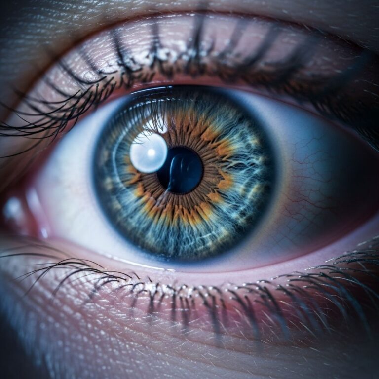
Facial discoid dermatosis (FDD) is a rare dermatological condition characterized by discrete, persistent pink-orange papulosquamous lesions confined exclusively to the face and occasionally the neck. First described in 2010 by Ko et al., FDD remains under-recognized due to its rarity, leading to frequent misdiagnosis as more common facial eruptions. Patients typically present with well-demarcated, annular plaques or papules exhibiting fine superficial scaling, which develop gradually over months to years and show remarkable resistance to conventional therapies.
The condition’s hallmark is its therapeutic recalcitrance, with lesions persisting despite trials of topical corticosteroids, antifungals, calcineurin inhibitors, and systemic agents like hydroxychloroquine and acitretin. This resistance not only causes cosmetic disfigurement but also significant psychological distress, particularly in younger patients or those with visible involvement on cheeks, forehead, or chin. Awareness of FDD is crucial for dermatologists to avoid unnecessary investigations and prolonged ineffective treatments.
Demographics
Limited case reports suggest FDD affects adults primarily, with onset ranging from adolescence to middle age. Reported cases include a 19-year-old male (Fitzpatrick skin type II), a 40-year-old female (type IV), a 44-year-old female (type III), and a 45-year-old woman. Cheeks are most commonly involved (93% of cases), followed by chin (69%) and forehead (38%). The condition appears stable over years, with no strong gender or ethnic predisposition identified due to paucity of data, though most reports are from diverse skin types.
- Age range: Adolescence to mid-40s in documented cases.
- Skin types: Reported across Fitzpatrick types II–IV; variation in darker skin types unknown.
- Gender: Both males and females affected equally in small series.
Causes
The aetiology of FDD remains unknown, with no identifiable infectious, autoimmune, or genetic triggers in reported cases. Histology consistently shows non-specific features like hyperkeratosis, parakeratosis, acanthosis, follicular plugging, and superficial dermal lymphocytic infiltrate, without evidence of fungi (negative PAS staining) or vasculopathy. Differential considerations like psoriasis or discoid lupus erythematosus (DLE) are ruled out by lack of response to targeted therapies and absent specific histological markers (e.g., no interface dermatitis for DLE).
One case noted abundant Demodex mites, prompting a trial of ivermectin and metronidazole for possible rosacea overlap, but without benefit. No systemic associations, such as depression treatment with fluoxetine, were causally linked. FDD may represent a distinct papulosquamous disorder limited to facial skin, possibly with subtle epidermal dysregulation.
Clinical Features
FDD manifests as multiple discrete, 0.5–2 cm annular or discoid plaques with pink-orange hue, mild scaling, and sharp margins, predominantly on the central face. Lesions are asymptomatic, lacking pruritus or pain, and remain stable without koebnerization or scarring. Progression is indolent, with gradual increase in number over years.
| Feature | Description |
|---|---|
| Appearance | Pink-orange papules/plaques, fine scale, annular |
| Location | Cheeks > chin > forehead; face/neck only |
| Symptoms | Asymptomatic |
| Duration | Months to years, persistent |
Photographs from cases depict orange-pink scaly discs on cheeks/forehead in lighter skin and erythematous plaques in darker tones.
Variation in Skin Types
Due to rarity, data on skin type variations is sparse. In Fitzpatrick type II (fair skin), lesions appear orange-pink with evident scale. Type III/IV show erythematous discs with subtle scaling, potentially mimicking seborrheic dermatitis. No hypopigmentation or keloidal changes reported in darker phototypes, but underreporting limits conclusions.
Complications
Primary complications are cosmetic disfigurement and emotional impact from persistent facial lesions. No scarring, ulceration, or secondary infection noted. One case developed type II pityriasis rubra pilaris (PRP) during follow-up, suggesting possible overlap with other papulosquamous disorders. Treatment side effects, like steroid atrophy from prolonged use, may arise due to misdiagnosis.
Diagnosis
Diagnosis is clinical, supported by biopsy to exclude mimics. Key histological findings include:
- Hyperkeratosis and focal parakeratosis
- Acanthosis and follicular plugging
- Superficial perivascular lymphocytic infiltrate
- Occasional subcorneal acantholysis or checkerboard orthokeratosis/parakeratosis
- Negative PAS for fungi
Skin scrapings for mycology are negative. Direct immunofluorescence is not featured but useful to rule out lupus.
Differential Diagnoses
Broad differentials for facial papulosquamous lesions include:
- Discoid lupus erythematosus (DLE): More indurated, scarring; interface dermatitis on biopsy.
- Psoriasis: Thicker plaques, responds to vitamin D/steroids.
- Tinea faciei: Annular, KOH/PAS positive.
- Seborrhoeic dermatitis: Greasy scale in seborrhoeic areas.
- Rosacea: Telangiectasia, papulopustular; Demodex-associated.
- Pityriasis rosea: Herald patch, truncal.
FDD is distinguished by facial exclusivity, resistance to therapy, and non-specific histology.
Treatment
No standardized treatment exists; all are empirical from case reports with poor response rates (47% failure). Commonly trialled:
Topical Therapies
- Potent/very potent corticosteroids (e.g., clobetasol, mometasone): No/minimal response.
- Calcineurin inhibitors (tacrolimus 0.1%): Partial in some.
- Vitamin D analogues (calcipotriol), combinations (betamethasone/calcipotriol): Partial success reported.
- Retinoids (tazarotene, tretinoin), antifungals (clotrimazole, miconazole), imiquimod: Ineffective.
Systemic Therapies
- Hydroxychloroquine (200–400 mg/day, 6–12 weeks): Failed in multiple cases.
- Acitretin (low-dose 25 mg/day) + topical calcipotriol/betamethasone: Successful in isolated reports.
- Antibiotics (doxycycline, oxytetracycline), antifungals (itraconazole), methotrexate, prednisolone: No benefit.
- Ustekinumab (IL-12/23 inhibitor): Recent success in one case.
Other
- NBUVB phototherapy, pulsed dye laser, intralesional steroids: Unsuccessful.
Partial response in 53% to tacrolimus/steroids or acitretin combos; ongoing trials recommended.
Prevention
No known preventive measures due to unclear aetiology. Avoid irritants and unnecessary topicals to prevent iatrogenic damage.
Outcome
Prognosis is guarded; lesions persist chronically with slow progression. Rare remissions with calcipotriol, low-dose acitretin, or ustekinumab reported, but most remain refractory. Long-term monitoring for papulosquamous evolution (e.g., PRP) advised.
Frequently Asked Questions
What is facial discoid dermatosis?
A rare, treatment-resistant condition with persistent pink-orange scaly plaques on the face.
Is FDD curable?
No definitive cure; partial responses to specific topicals/systemics in select cases.
How is FDD diagnosed?
Clinical + biopsy excluding differentials like DLE, psoriasis, tinea.
What does FDD look like on different skin types?
Pink-orange in light skin; erythematous in medium tones; data limited for dark skin.
Does FDD scar?
No, lesions are non-scarring and asymptomatic.
References
- Facial discoid dermatosis: A cosmetically disfiguring and treatment-resistant facial dermatosis — Antony C, et al. 2022-04-15. https://pmc.ncbi.nlm.nih.gov/articles/PMC9060026/
- Facial Discoid Dermatosis: An Enigmatic Disease — Acta Dermosifiliogr. 2022. https://www.actasdermo.org/es-facial-discoid-dermatosis-an-enigmatic-articulo-S1578219021002912
- Facial discoid dermatosis — DermNet NZ. Recent update. https://dermnetnz.org/topics/facial-discoid-dermatosis
Similar Articles


Read full bio of Sneha Tete