Flap Surgery and Reconstructive Microsurgery
Advanced surgical techniques for restoring form and function through tissue reconstruction.

Flap Surgery and Reconstructive Microsurgery: Restoring Form and Function
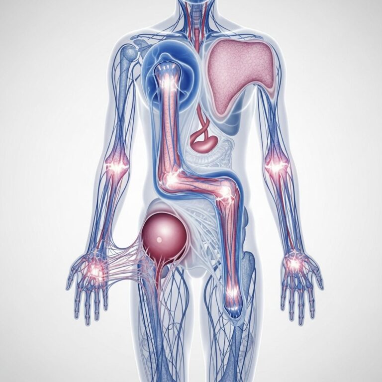
Flap surgery and reconstructive microsurgery represent some of the most advanced techniques in modern medicine, offering solutions for patients who have experienced significant tissue loss due to injury, disease, or surgical treatment. These sophisticated procedures involve transferring healthy tissue from one area of the body to another, or using specialized surgical techniques to reconnect blood vessels at the microscopic level. Whether you have suffered a traumatic injury, undergone cancer treatment, or deal with a congenital condition, flap surgery and reconstructive microsurgery can help restore both form and function to affected areas.
Understanding Flap Surgery
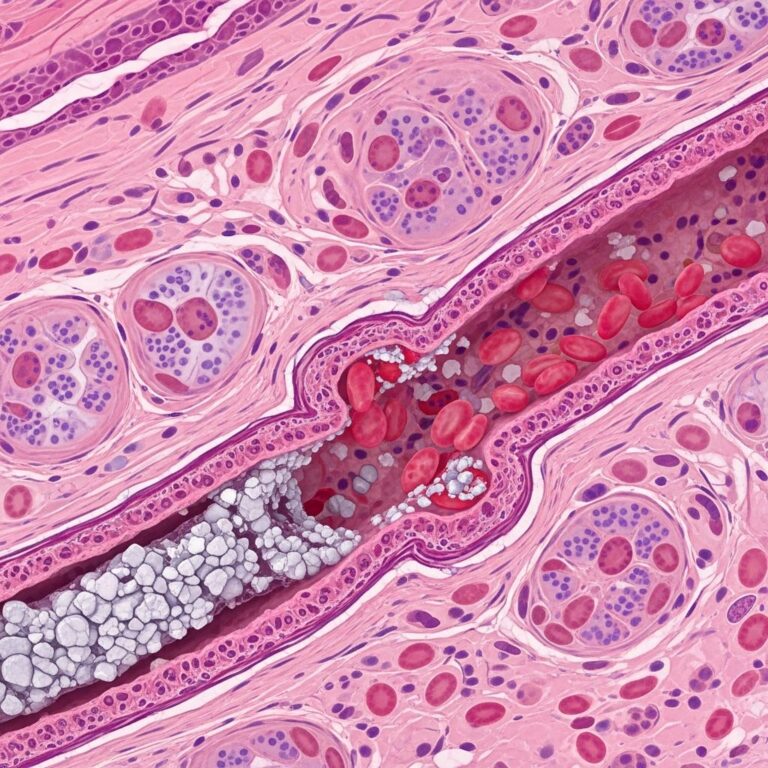
Flap surgery is a reconstructive procedure that involves moving living tissue from one part of the body to another, maintaining the tissue’s blood supply so it can survive and heal in its new location. The tissue transferred, known as a flap, can include skin, fat, muscle, bone, or a combination of these structures. The key advantage of flap surgery is that the tissue retains its own blood supply through intact blood vessels, allowing for superior healing and long-term viability compared to simpler grafting techniques.
The use of flaps in reconstructive surgery dates back decades, but modern advances have significantly improved outcomes and expanded the range of conditions that can be treated. Surgeons now can repair complex defects involving multiple tissue types with remarkable precision and aesthetic results.
Types of Flaps
Several types of flaps are available depending on the nature and location of the defect requiring repair:
- Local Flaps: Tissue is moved from an area adjacent to the defect, keeping the blood supply connected through a narrow bridge of tissue called a pedicle. Local tissue flaps are commonly used in facial reconstructive surgery because they provide skin texture and color that closely matches the surrounding area.
- Regional Flaps: These flaps are obtained from tissues near the defect but farther away than local flaps. The blood supply is maintained through a pedicle that connects the flap to its original location.
- Free Flaps: The tissue is completely detached from its original location and transferred to the defect site, where microsurgical techniques are used to reconnect its blood vessels to new blood vessels at the recipient site. Free flaps offer greater flexibility in reconstruction because the surgeon can choose tissue from distant areas of the body that best match the requirements.
The Role of Reconstructive Microsurgery
Reconstructive microsurgery is the specialized application of microsurgical techniques to reconstruct complex tissue defects. This approach has revolutionized the field of plastic and reconstructive surgery, particularly in treating head and neck cancers, severe facial injuries, breast reconstruction, and other challenging cases.
Microsurgical Techniques and Applications
Microsurgery involves operating under magnification, typically using operating microscopes that provide magnification of 4 to 25 times the normal view. This allows surgeons to work with structures as small as blood vessels with diameters of less than one millimeter. When performing free flap surgery, microsurgeons must carefully identify recipient blood vessels and perform precise anastomoses—connections between the flap’s blood vessels and the recipient vessels.
At high-volume centers such as Johns Hopkins and other major medical institutions, surgeons perform complex microvascular reconstructions routinely. These centers perform approximately 200 or more free flap reconstructions annually, sometimes performing four to six procedures per week. This high volume contributes to superior outcomes, as experience significantly impacts success rates.
One innovative advancement in flap design comes from computer-assisted surgery applications. These tools help surgeons create and plan local tissue flaps in facial reconstructive surgery by providing three-dimensional reconstructed images of the patient’s anatomy. Surgeons can view multiple flap designs on a 3D model, receive tension and strain feedback, and make informed edits to optimize the surgical plan before making the initial incision. This technology has the potential to improve outcomes and reduce the learning curve for less experienced surgeons.
Common Indications for Flap Surgery
Flap surgery and reconstructive microsurgery are used to treat a wide variety of conditions:
- Cancer Reconstruction: Following surgical removal of head and neck cancers, flap surgery can restore structure and function to areas including the jawbone, throat, and facial features.
- Traumatic Injuries: Severe burns, lacerations, crush injuries, and other trauma may result in extensive tissue loss requiring flap reconstruction.
- Breast Reconstruction: After mastectomy for breast cancer, free flap techniques such as the DIEP (Deep Inferior Epigastric Perforator) flap provide natural-looking and feeling breast reconstruction using the patient’s own tissue.
- Osteoradionecrosis: Patients who have undergone radiation therapy for head and neck cancer may develop bone necrosis (death) months or years after treatment. Limited free flap procedures can address this complication with shorter hospital stays and recovery periods compared to traditional extensive reconstructions.
- Congenital Defects: Birth defects affecting the head, neck, or other areas may benefit from flap reconstruction.
The DIEP Flap for Breast Reconstruction
The DIEP (Deep Inferior Epigastric Perforator) flap represents an important advancement in breast reconstruction following mastectomy. This procedure uses tissue from the lower abdomen, including skin and fat but not the abdominal muscle. During the procedure, an incision is made from hip to hip, and the skin, fat, and blood vessels that will be used for reconstruction are carefully separated from the abdominal muscle. The tissue is then removed and transplanted into the breast pocket, where the blood vessels are connected to blood vessels in the chest to provide adequate circulation for the reconstructed breast.
The DIEP flap offers significant advantages over older techniques such as the TRAM (Transverse Rectus Abdominis Muscle) flap. Because the abdominal muscle is preserved, patients experience fewer complications at the donor site, including reduced risk of hernia and abdominal bulging. Studies show that abdominal hernia risk is typically in the four to five percent range with DIEP flaps, much lower than the rates associated with TRAM flaps. Additionally, the reconstructed breast has excellent shape and natural appearance.
Enhanced Recovery After Surgery Protocols
Modern flap surgery increasingly incorporates enhanced recovery after surgery (ERAS) pathways, which optimize patient outcomes through evidence-based perioperative protocols. While well-established in other surgical specialties, ERAS pathways are relatively new to plastic and reconstructive surgery but show promising results.
Multimodal Pain Management
Effective pain control is essential for patient comfort and recovery. Preoperative analgesic regimens typically include celecoxib, gabapentin, and oral acetaminophen. Intraoperative regional anesthesia blocks, such as transversus abdominis plane blocks performed by the anesthesia team during microsurgical anastomosis, provide excellent postoperative pain control without extending operative time when coordinated with the surgical team.
Thromboembolism Prevention
Patients undergoing free flap surgery, particularly for breast reconstruction, face increased risk for blood clots due to factors such as advanced age, prolonged operative time, history of malignancy, and postoperative immobility. Risk stratification helps identify high-risk patients who benefit from extended low molecular weight heparin treatment following discharge. Current evidence-based recommendations suggest pharmacologic anticoagulation should continue for at least seven days postoperatively in high-risk patients.
Early Feeding
Evidence from other surgical specialties demonstrates that early feeding following surgery reduces complications and mortality without increasing the risk of flap compromise. Modern protocols now include sips of water or ice chips the evening following surgery, with formal diet advancement as tolerated beginning the morning after surgery. This approach maintains nutritional support while preserving the ability to return to the operating room rapidly should flap concerns develop.
Recovery and Outcomes
Recovery from flap surgery varies depending on the extent of the procedure and the type of flap used. Patients typically remain hospitalized for several days following surgery, with discharge occurring once adequate healing has begun and pain is controlled. Some procedures, particularly limited free flap reconstructions for conditions such as osteoradionecrosis, may allow discharge within just a couple of days.
Physical activity should be gradually resumed under the guidance of the surgical team. Vigorous activity and heavy lifting should be avoided for several weeks to allow the flap to establish adequate blood supply and the surgical sites to heal completely.
Outcomes following flap surgery are generally excellent when performed by experienced surgeons at high-volume centers. Success rates for free flap surgery exceed 95 percent at major academic medical centers. Patients achieve restoration of form and function, with improvements in both appearance and ability to eat, speak, and breathe normally when applicable.
Technological Advances in Flap Surgery
Recent technological innovations are enhancing flap surgery planning and execution. Three-dimensional imaging and computer-assisted design platforms allow surgeons to visualize anatomy in unprecedented detail and plan optimal flap designs preoperatively. These tools provide immediate feedback regarding tissue tension and strain, enabling surgeons to refine their surgical approach before the procedure begins. Such technology is particularly valuable for novice and less experienced surgeons, potentially accelerating the learning curve for challenging flap-design decisions that traditionally required many years of operating experience to master.
Frequently Asked Questions
What is the difference between a skin graft and a flap?
Skin grafts involve transplanting skin without its own blood supply, relying instead on blood from the recipient site. Flaps include living tissue with an intact blood supply, resulting in better healing and more durable reconstruction. Flaps are preferred for complex defects involving multiple tissue types.
How long does flap surgery take?
Operative time varies significantly depending on the complexity of the reconstruction. Local and regional flap procedures may take one to three hours, while free flap surgery typically requires four to eight hours or longer. The surgeon’s experience and the complexity of the defect being repaired are primary factors affecting duration.
What are the risks of flap surgery?
Potential complications include flap failure or partial loss due to inadequate blood supply, infection, bleeding, and blood clots. Donor site complications may include hernia, weakness, or nerve injury depending on the flap type and location. However, complication rates are relatively low at experienced centers.
Is flap surgery covered by insurance?
Most insurance plans cover flap surgery when it is medically necessary for reconstruction following cancer treatment, trauma, or significant injury. Cosmetic flap procedures typically are not covered. Patients should discuss coverage with their insurance provider and the surgeon’s billing department.
Can flap surgery improve appearance?
Yes, one of the primary goals of flap surgery is to restore both form and function. When properly planned and executed, flap surgery results in reconstruction that is close to unnoticeable. The surgeon carefully considers aesthetic outcomes alongside functional restoration.
How long does recovery take?
Initial recovery typically takes two to four weeks, with return to light activities possible after this period. Full recovery, including return to all normal activities, may require two to three months or longer. The timeline depends on the extent of surgery and individual healing.
Will I have scars after flap surgery?
Yes, flap surgery results in scars at both the donor site (where tissue is taken) and recipient site (where tissue is placed). However, experienced surgeons place incisions strategically to minimize visible scarring. Scars typically fade and become less noticeable over 12 to 18 months.
References
- Flapp: A Platform for 3D Local Soft-tissue Flap Design for Facial Reconstructive Surgery — Johns Hopkins University, Department of Biomedical Engineering. 2023. https://www.bme.jhu.edu/hello-world/flapp-a-platform-for-3d-local-soft-tissue-flap-design-for-facial-reconstructive-surgery/
- Implementing Our Microsurgical Breast Reconstruction Enhanced Recovery After Surgery Pathway — National Center for Biotechnology Information, U.S. National Library of Medicine. 2019. https://pmc.ncbi.nlm.nih.gov/articles/PMC6382235/
- Microvascular Head and Neck Reconstructive Surgery — Johns Hopkins Medicine. 2023. https://www.youtube.com/watch?v=W0Pw9UjKxWE
- Breast Reconstruction Surgery – DIEP Flap — Johns Hopkins Medicine. 2010. https://www.youtube.com/watch?v=tYEt1Pw6FNU
- Understanding the Breast Reconstructive Ladder — Johns Hopkins Medicine. 2020. https://www.youtube.com/watch?v=00RdG7EjjMs
Similar Articles


Read full bio of medha deb