Fracture Blister: Causes, Risks, And Management Guide
Understanding fracture blisters: causes, diagnosis, management, and complications of these tense fluid-filled blisters overlying acute fractures.

A
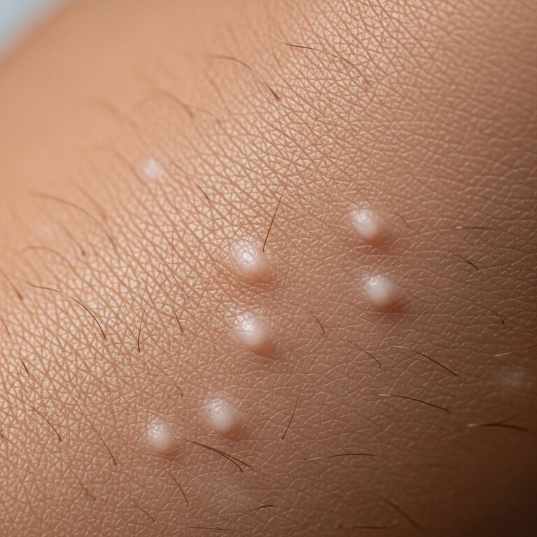
fracture blister
is a tense, fluid-filled blister that develops on the skin directly overlying an acute bony fracture. These blisters resemble those seen in second-degree burns but arise from mechanical trauma rather than heat. They complicate approximately3% of fractures
requiring hospital treatment, particularly in areas with thin subcutaneous tissue.Fracture blisters pose significant challenges in orthopaedic management, as they overlie potential surgical incision sites, delay stabilization, and increase infection risks. Early recognition and appropriate care are essential to optimize healing and prevent complications.
Demographics
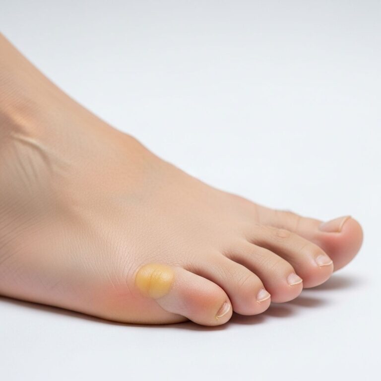
Fracture blisters occur in
2.9–3%
of hospitalized fracture patients, with higher incidence in high-energy trauma cases. They predominantly affect adults involved in motor vehicle accidents, falls from height (average 18 feet), or pedestrian injuries. Children and elderly patients are less commonly affected due to differences in skin adherence and injury mechanisms.- Prevalence by site: Ankle (most common, up to 25% in some series), distal tibia, wrist, elbow, foot.
- Risk elevation: Males in high-impact scenarios; patients with comorbidities like diabetes or vascular disease.
Causes
Fracture blisters result from a combination of mechanical shearing forces and post-traumatic physiological changes. The primary mechanism involves
large strains on the skin
during fracture deformation, creating a cleavage injury at the dermo-epidermal junction.Pathophysiology
When bone angulates during fracture, tightly adherent skin (with minimal subcutaneous fat or muscle) experiences shear stress. This strain—calculated as up to
152%
in models—separates epidermal layers (clear blisters) or the epidermis from dermis (hemorrhagic blisters).Additional contributors include:
- Post-traumatic edema: Increased interstitial pressure disrupts epidermal cohesion, facilitating fluid accumulation.
- Venous/lymphatic compromise: Thrombosis and hypoxia lead to epidermal necrosis.
- Compartment pressure: Elevated pressures in limb compartments drive fluid into subepidermal spaces.
| Type | Fluid | Separation Level | Healing Time | Severity |
|---|---|---|---|---|
| Serous (Clear) | Clear fluid | Intra-epidermal | ~12 days | Milder |
| Hemorrhagic | Blood-tinged | Dermo-epidermal junction | ~16 days | More severe |
Blisters form rapidly: as early as
6 hours
post-injury, peaking at24–48 hours
.Risk Factors
- Anatomical: Ankle, wrist, elbow, foot, distal tibia (thin skin, tight adherence).
- Injury-related: High-energy trauma, grade I/II open tibia fractures, crush injuries.
- Patient factors: Peripheral vascular disease, diabetes, smoking, hypertension, alcoholism, lymphatic obstruction.
Diagnosis
Diagnosis is primarily
clinical
, based on tense vesicles or bullae over markedly swollen skin directly above a confirmed fracture. Imaging (X-ray/CT) verifies the underlying fracture.Key features:
- Tense blister with clear or hemorrhagic fluid.
- Surrounding ecchymosis and edema.
- No history of thermal injury.
If uncertain,
skin biopsy
reveals:- Subepidermal split with minimal inflammation.
- Dermal elastin damage.
- No significant leukocyte infiltrate (vs. infection).
Differentiate from friction blisters (epidermal only) or burns (thermal history, deeper necrosis).
Prevention
While not always preventable, strategies focus on minimizing swelling and shear:
- Immediate post-injury: Strict elevation, icing (if not contraindicated), compression wraps (avoid over-tight splints).
- Early intervention: Stabilize fracture before blister formation (prophylactic pinning if high-risk).
- Risk modification: Optimize comorbidities (e.g., smoking cessation, glycemic control).
- Avoid early tight casting; monitor high-risk sites closely.
Management
No universal consensus exists, but most experts recommend
non-operative initial approach
to allow resolution.Conservative Care
- Do not deroof or aspirate: Risks infection; intact skin barrier protects underlying fracture.
- Wound care: Non-adherent dressings, topical antibiotics if ruptured; daily inspection.
- Elevation/immobilization: Above heart level to reduce edema; loose splints.
- Pain/swelling control: NSAIDs, analgesics; monitor for compartment syndrome.
Surgery delayed
until re-epithelialization
(10–21 days); serous blisters resolve faster than hemorrhagic.Surgical Considerations
- Incise away from blister site.
- If infected/large: Debride post-resolution.
- Early fixation in select cases (e.g., external fixator).
| Phase | Days Post-Injury | Actions |
|---|---|---|
| Acute | 0–3 | Elevate, observe, loose splint |
| Subacute | 3–10 | Dressings, monitor healing |
| Definitive | 10–21 | Surgery if needed |
Complications
Fracture blisters significantly worsen outcomes:
- Delayed surgery: Average 12–16 days, increasing non-union risk.
- Infection: Wound dehiscence, osteomyelitis (up to 20% in complicated cases).
- Chronic ulcers/scarring: Especially hemorrhagic types.
- Prolonged hospitalization: Mean increase of 7–10 days.
High-risk patients (e.g., diabetics) face amplified complications; vigilant monitoring is critical.
Frequently Asked Questions (FAQs)
Q: How soon do fracture blisters appear?
A: As early as 6 hours, but most within 24–48 hours post-fracture.
Q: Should I pop a fracture blister?
A: No—popping increases infection risk. Keep intact and seek medical advice.
Q: Can fracture blisters occur after surgery?
A: Yes, after osteotomies or realignments causing skin shear.
Q: How long until surgery is safe?
A: Wait for re-epithelialization: 12 days (serous), 16 days (hemorrhagic).
Q: Are fracture blisters contagious?
A: No, they are sterile trauma responses unless secondarily infected.
References
- Fracture Blisters — Varela R, et al. PMC – NIH. 2011-04-15. https://pmc.ncbi.nlm.nih.gov/articles/PMC3088393/
- Fracture Blisters — DermNet NZ. 2023-01-01. https://dermnetnz.org/topics/fracture-blister
- Fracture Blister — Wikipedia (sourced from peer-reviewed refs). 2024-06-12. https://en.wikipedia.org/wiki/Fracture_blister
- Fracture Blisters: A Commonly Asked Patient Question — Ohio Foot and Ankle Surgeon. 2023-05-20. https://www.ohiofootandanklesurgeon.com/blog/fracture-blisters-a-commonly-asked-patient-question
- Fracture Blisters: Causes, Treatment, and How Long They Last — Healthline (medically reviewed). 2018-03-21. https://www.healthline.com/health/fracture-blisters
Similar Articles


Read full bio of Sneha Tete