Fuchs’ Dystrophy: 3 Stages, Symptoms, And Treatments
Discover the causes, stages, symptoms, and latest treatments for Fuchs' dystrophy, a progressive corneal condition affecting vision clarity.

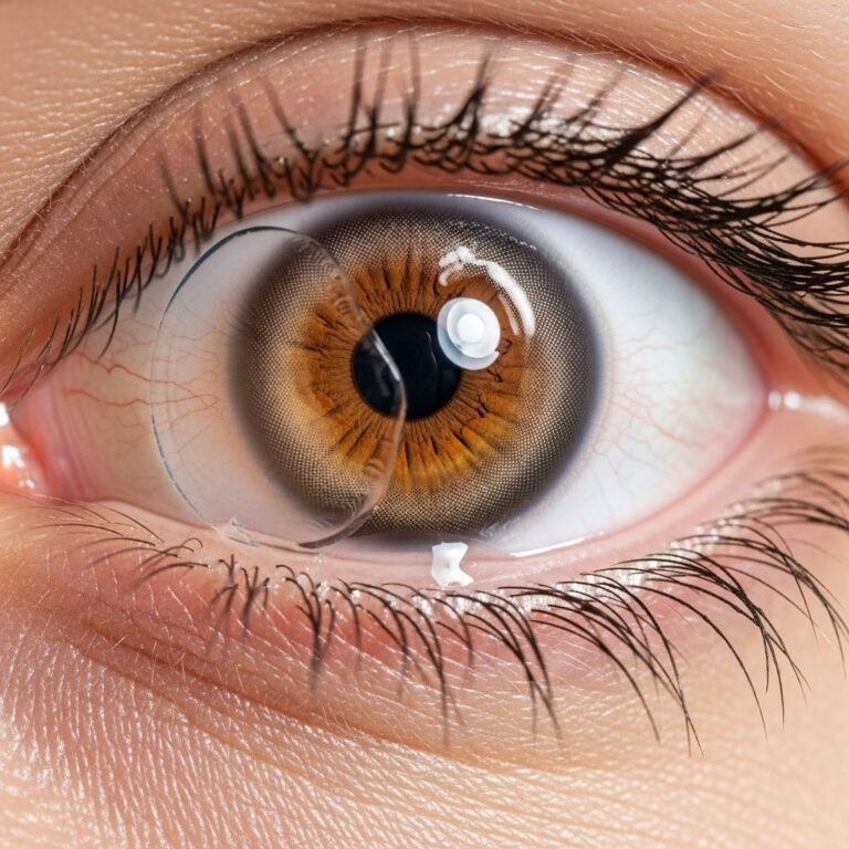
Fuchs’ dystrophy, also known as Fuchs’ endothelial dystrophy, is a progressive eye condition that primarily affects the cornea’s innermost layer. This disease leads to gradual vision deterioration due to the malfunction and loss of endothelial cells, which are crucial for maintaining corneal clarity by pumping out excess fluid. Over time, it can significantly impair daily activities, but early detection and appropriate interventions can help preserve vision.
Understanding the Cornea and Its Vital Role
The cornea is the clear, dome-shaped front surface of the eye that helps focus light onto the retina. It consists of five layers: epithelium, Bowman’s layer, stroma, Descemet’s membrane, and endothelium. The endothelium, a single layer of cells on the inner surface, acts as a pump to remove fluid from the stroma, keeping the cornea thin and transparent. In Fuchs’ dystrophy, these cells drop in number and function, causing fluid buildup, swelling (edema), and loss of transparency.
Endothelial cells do not regenerate, so any loss is permanent. Healthy adults have about 2,500 to 3,000 cells per square millimeter at birth, decreasing naturally with age to around 2,000 by age 40. In Fuchs’ dystrophy, this decline accelerates, often exacerbated by genetic factors.
Genetic Roots and Risk Factors
Fuchs’ dystrophy is primarily genetic, inherited in an autosomal dominant pattern with variable penetrance, meaning not all carriers show symptoms equally. It can also occur sporadically without family history. Women are affected more frequently than men, possibly due to hormonal influences or longer lifespan.
The condition typically manifests after age 50, though guttata—small collagen excrescences on Descemet’s membrane—may appear earlier. Risk increases with age, and environmental factors like humid climates may worsen symptoms by hindering fluid evaporation from the cornea. Unlike inflammatory conditions, Fuchs’ is non-inflammatory, stemming from intrinsic cellular defects.
Progressive Stages of the Disease
Fuchs’ dystrophy advances slowly through distinct phases, each marked by worsening corneal changes and vision impact.
- Early Stage (Guttae Formation): Abnormal bumps called guttata develop on Descemet’s membrane, disrupting the endothelium’s smooth surface. Vision remains relatively normal, but subtle glare or halos around lights may emerge, especially at night.
- Intermediate Stage (Corneal Edema): Endothelial cell loss reduces pumping efficiency, leading to stromal edema. Vision blurs primarily in the morning due to overnight fluid accumulation, improving as eyes open and evaporate excess during the day. Symptoms intensify in humid weather.
- Advanced Stage (Bullous Keratopathy): Persistent edema reaches the epithelium, forming painful blisters (bullae). Ruptured bullae cause epithelial defects, pain, sensitivity, and risk of infection. Chronic changes lead to subepithelial fibrosis, scarring, haze, and vascularization, severely clouding vision.
This progression can span decades, with some individuals remaining asymptomatic until late stages.
Recognizing Key Symptoms
Symptoms evolve with disease advancement. Initial signs include:
- Glare and halos around lights, particularly at night.
- Mild vision reduction and poor contrast sensitivity.
- Fluctuating blurry vision, worse upon waking.
As edema persists, patients report constant haziness, photophobia (light sensitivity), foreign body sensation, and eye pain from bullae rupture. Night driving becomes hazardous due to light scatter.
Importantly, Fuchs’ symptoms differ from dry eye: blurriness does not improve with blinking or tears, distinguishing it diagnostically.
Diagnostic Approaches
Diagnosis begins with a comprehensive eye exam. Key methods include:
| Method | Purpose | Findings in Fuchs’ |
|---|---|---|
| Slit-Lamp Biomicroscopy | Examines corneal layers | Guttae, endothelial folds, stromal haze, bullae |
| Specular Microscopy | Counts endothelial cells | Low density (<1,000 cells/mm²), pleomorphism |
| Pachymetry | Measures thickness | Increased central thickness (>600 µm) |
| Corneal Topography | Assesses surface | Irregular astigmatism from edema |
Early detection via specular microscopy is crucial, as cell counts predict progression. Fluorescein staining reveals microcysts or defects.
Management and Treatment Options
Treatment is stage-dependent, focusing on symptom relief and halting progression.
Conservative Measures
- Hypertonic Saline Drops/Ointments: Draw out fluid, reducing morning edema. Use before bed and upon waking.
- Bandage Contact Lenses: Protect bullae, alleviate pain in advanced cases.
- Avoid Eye Rubbing: Prevents endothelial trauma.
Surgical Interventions
When vision loss impacts quality of life (typically <20/40 acuity or severe pain), surgery replaces dysfunctional endothelium.
- Endothelial Keratoplasty (EK): Preferred over full-thickness transplant. Includes Descemet’s Stripping Automated Endothelial Keratoplasty (DSAEK) and Descemet’s Membrane Endothelial Keratoplasty (DMEK). DMEK offers faster recovery and better vision.
- Penetrating Keratoplasty (PK): Rarely used now, for severe scarring.
Success rates exceed 90%, with low rejection risk due to minimal donor tissue. Newer techniques like DMEK yield outcomes close to natural corneas.
Living with Fuchs’ Dystrophy
Regular monitoring by a corneal specialist is essential. Lifestyle adjustments include using humidifiers sparingly, artificial tears for comfort (though not curative), and sunglasses for glare. Genetic counseling may benefit families.
Research explores gene therapy and endothelial cell injections, but transplants remain gold standard.
Frequently Asked Questions (FAQs)
Is Fuchs’ dystrophy hereditary?
Yes, often autosomal dominant, but variable expression means family members may vary in severity.
Can Fuchs’ dystrophy affect both eyes?
Typically bilateral, though one eye may progress faster.
How fast does Fuchs’ dystrophy progress?
Over decades; early stages may be asymptomatic for years.
Does laser surgery help Fuchs’ dystrophy?
No, it does not address endothelial dysfunction; transplants are required.
What is the prognosis after corneal transplant?
Excellent, with 95% graft survival at 5 years and restored vision.
References
- Fuchs’ Endothelial Dystrophy — EyeWiki (American Academy of Ophthalmology). 2023-10-15. https://eyewiki.org/Fuchs%E2%80%99_Endothelial_Dystrophy
- An Introduction to Fuchs’ Dystrophy for Patients — Corneal Dystrophy Foundation. 2024-01-12. https://www.cornealdystrophyfoundation.org/an-introduction-to-fuchs-dystrophy-for-patients/
- Fuchs’ Corneal Dystrophy — Dr. McDevitt EyeCare. 2023-05-20. https://drmcdevitteyecare.com/articles/general/418116-fuchs-corneal-dystrophy
- Fuchs’ Dystrophy – Symptoms, Causes & Treatment — Lions Eye Institute. 2024-02-10. https://www.lei.org.au/services/eye-health-information/fuchs-dystrophy/
- What is Fuchs’ Dystrophy? — Price Vision Group. 2023-11-08. https://pricevisiongroup.com/conditions/fuchs-dystrophy/
- Fuchs’ Dystrophy: What It Is, Symptoms & Treatment — Cleveland Clinic. 2024-08-22. https://my.clevelandclinic.org/health/diseases/23438-fuchs-dystrophy
Similar Articles


Read full bio of medha deb