Generalized Eruptive Keratoacanthomas: Diagnosis And Treatment
Rare skin disorder with hundreds of keratoacanthoma-like papules causing significant morbidity and treatment challenges.

Introduction
Generalised eruptive keratoacanthoma of Grzybowski, commonly referred to as Grzybowski syndrome, represents a rare and distinct variant of keratoacanthoma. This condition is defined by the explosive or gradual onset of hundreds to thousands of small, keratoacanthoma-like papules disseminated across the skin and mucous membranes. First documented in 1950 by Polish dermatologist Jan Grzybowski, fewer than 50 cases have been reported in medical literature to date, underscoring its extreme rarity.
Unlike solitary keratoacanthomas, which are relatively common and often self-resolve, Grzybowski syndrome manifests as a chronic, progressive eruption that poses substantial cosmetic and functional challenges. Lesions typically measure 1–5 mm in diameter, appearing as pruritic, umbilicated papules with central keratotic plugs. These eruptions can profoundly impact quality of life due to their sheer volume, itchiness, and potential for scarring.
The syndrome’s hallmark is its generalised distribution, favouring sun-exposed regions while also affecting intertriginous areas and oral mucosa. Although benign in terms of malignant transformation, the disease’s persistence and resistance to therapy make it a formidable clinical entity for dermatologists.
Demographics
Generalised eruptive keratoacanthomas predominantly affect adults in their fifth to seventh decades of life, with cases reported between ages 40 and 70. There is no clear gender predilection, as both males and females are equally represented in documented cases.
The condition occurs sporadically without a familial pattern or hereditary component, distinguishing it from other multiple keratoacanthoma syndromes like Ferguson-Smith type, which follows an autosomal dominant inheritance. Skin phototypes span all categories, with no racial or ethnic predisposition identified, though most reports originate from Caucasian populations due to reporting biases in dermatological literature.
Comorbidities noted in some patients include internal malignancies (e.g., breast cancer), hypertension, psoriasis, and chronic kidney disease, though no definitive causal links exist. Exposure to chemical carcinogens or UV radiation has been speculated but not proven.
Causes
The precise aetiology of generalised eruptive keratoacanthomas remains elusive. Unlike solitary keratoacanthomas occasionally linked to human papillomavirus (HPV), no such association has been confirmed in the eruptive variant. Potential predisposing factors include:
- Chronic UV exposure, given the predilection for sun-damaged areas like the face and upper trunk.
- Immune dysregulation, such as interleukin-2 deficiency or other immunocompromised states.
- Chemical carcinogen exposure, though unproven.
- Genetic mutations in keratinocyte proliferation pathways, inferred from histological similarities to squamous proliferations.
No hereditary pattern is observed, and the abrupt onset suggests a possible somatic trigger in susceptible individuals. Recent case reports highlight associations with tamoxifen use or prior malignancies, but these are anecdotal.
Clinical Features
The eruption can onset suddenly or progressively over weeks to months, yielding hundreds to thousands of lesions. Key characteristics include:
- Size and Morphology: Small papules (1–5 mm) that are skin-coloured to erythematous, umbilicated, with a central hyperkeratotic plug.
- Symptoms: Intense pruritus, occasional pain from ulceration.
- Distribution: Predominantly sun-exposed sites (face, upper trunk), intertriginous areas (axillae, groin), and mucous membranes (tongue, buccal mucosa, larynx). Palms and soles are characteristically spared.
Facial involvement may be florid, with periorbital confluence creating a ‘mask-like’ or ‘sign of Zorro’ appearance, potentially leading to ectropion. Lesions evolve from papules to crateriform nodules, sometimes ulcerating before forming atrophic scars upon partial involution.
Variation in Skin Types
While most cases are described in lighter skin phototypes (Fitzpatrick I–III), generalised eruptive keratoacanthomas have been documented across all skin types. In darker phototypes (IV–VI), lesions may appear hyperpigmented rather than erythematous, with similar morphology and distribution.
Pruritus remains universal, but scarring may be more pronounced in higher phototypes due to post-inflammatory hyperpigmentation. Mucosal involvement does not vary by skin type, and diagnostic challenges in pigmented skin arise from mimicry with other papular eruptions like prurigo nodularis.
Complications
This chronic condition inflicts considerable morbidity:
- Cosmetic disfigurement: Extensive facial scarring and mask-like facies.
- Functional impairment: Ectropion, oral discomfort, laryngeal involvement affecting speech/swallowing.
- Psychological impact: Severe pruritus and visible lesions leading to social withdrawal.
- Secondary issues: Infection from excoriation, koebnerisation from trauma.
Malignant transformation to squamous cell carcinoma is exceptionally rare, with no confirmed metastases reported.
Diagnosis
Diagnosis relies on characteristic clinical presentation corroborated by histopathology. Typical histology mirrors solitary keratoacanthoma: crateriform architecture, acanthosis, papillomatosis, atypical keratinocytes, central keratin plug, and inflammatory infiltrate.
Biopsy is essential to exclude squamous cell carcinoma, which shares overlapping features. Dermoscopy may reveal central keratin plugs with peripheral vessels. No specific laboratory markers exist.
Differential Diagnoses
| Condition | Key Distinguishing Features |
|---|---|
| Ferguson-Smith multiple keratoacanthomas | Autosomal dominant, adolescent onset, fewer lesions, spontaneous regression. |
| Solitary keratoacanthoma | Single lesion, self-resolves in 3–6 months. |
| Squamous cell carcinoma | Aggressive growth, atypia on biopsy, potential metastasis. |
| Prurigo nodularis | Larger nodules, no keratin plugs, intensely pruritic without umbilication. |
| Multiple trichoepitheliomas | Symmetrical facial distribution, no mucous membrane involvement. |
Treatment
Treatment remains challenging due to lesion multiplicity and recurrence risk. No curative option exists; management is palliative.
Oral Retinoids (Most Effective)
Acitretin or isotretinoin (0.5–1 mg/kg/day) induces regression in many cases, though relapse occurs upon discontinuation. A case report noted complete clearance with isotretinoin after 2 years.
Surgical and Physical Modalities
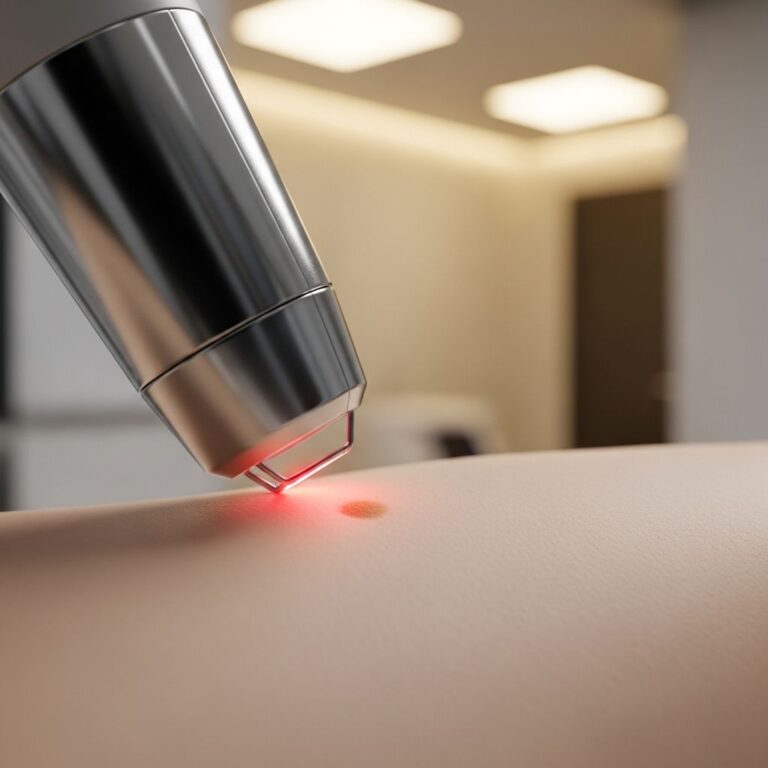
- Excision or Mohs surgery for localised clusters, impractical for widespread disease.
- Cryotherapy, electrodesiccation, laser ablation – risk koebnerisation.
Topical Therapies
- 5-Fluorouracil, imiquimod, retinoids – limited efficacy for numerous lesions.
Intralesional Therapies
- 5-Fluorouracil, bleomycin, steroids – feasible for accessible sites.
Other Systemic Therapies
- Methotrexate, interferon-alpha, ranitidine – anecdotal success.
Multimodal approaches are often required, with patient selection based on lesion burden and comorbidities.
Outcome
The disease is typically chronic and progressive, with unsatisfactory response to therapy. Partial remissions occur, but new crops emerge. Long-term retinoid maintenance may control outbreaks. Scarring persists lifelong, but malignancy risk is negligible. Prognosis focuses on symptom control and quality-of-life preservation.
Frequently Asked Questions
What causes generalised eruptive keratoacanthomas?
The exact cause is unknown, but associations with UV exposure, immune factors, and chemical triggers are proposed without confirmation.
Is it cancerous?
No malignant transformation or metastasis has been reported; it is considered benign despite histological similarities to squamous cell carcinoma.
Can it be cured?
No cure exists; oral retinoids offer the best control, but relapses are common.
Does it affect mucous membranes?
Yes, tongue, buccal mucosa, and larynx can be involved.
Is it hereditary?
No, it is sporadic without familial inheritance.
References
- Generalised eruptive keratoacanthomas — DermNet NZ. Accessed 2026. https://dermnetnz.org/topics/generalised-eruptive-keratoacanthomas
- Generalized eruptive keratoacanthomas — eScholarship, University of California. 2005. https://escholarship.org/uc/item/6b8870c5
- Squamous Cell Cancer – Generalized Eruptive Keratoacanthomas — Perris Dermatology (medically reviewed). 2011-01-16. https://perridermatology.com/dr-perris-blog/squamous-cell-cancer-generalized-eruptive-keratoacanthomas/
- Generalized Eruptive Keratoacanthomas of Grzybowski Treated with Isotretinoin — Journal of Drugs in Dermatology. 2008-11. https://jddonline.com/articles/generalized-eruptive-keratoacanthomas-of-grzybowskitreated-with-isotretinoin-S1545961608P1069X
- Generalized Eruptive Keratoacanthomas of Grzybowski and the Association with Chronic Kidney Disease — SKIN The Journal of Cutaneous Medicine. Recent. https://skin.dermsquared.com/skin/article/view/2169
Similar Articles


Read full bio of Sneha Tete