Glomus Tumour: 4 Types, Diagnosis, And Treatment
Rare, painful benign skin tumours arising from glomus cells, often under nails, with surgical cure.

A
glomus tumour
is a rare, typically benign neoplasm originating from the glomus cells within the Sucquet-Hoyer canal of the glomus body, a specialised arteriovenous anastomosis in the skin involved in thermoregulation. These tumours most commonly present as intensely painful nodules, particularly under the nail bed (subungual) or in the fingers and palms of young adults.What is a glomus tumour?
The glomus body is a temperature-sensitive structure composed of modified smooth muscle cells surrounding capillary loops, primarily located in the dermis of acral sites like fingers, toes, and palms. It regulates cutaneous blood flow: contracting in cold to conserve heat and dilating in warmth to promote dissipation.
Glomus tumours arise from proliferation of these glomus cells, forming well-circumscribed nodules around vascular channels. They are usually <1 cm in size but cause disproportionate pain due to high nerve density in the area. While 75% are solitary and sporadic, multiple lesions suggest inherited forms like glomuvenous malformations. Extracutaneous sites (e.g., GI tract, lung) are exceptional.
Who gets glomus tumours?
Glomus tumours affect adults aged 20–40 years equally in both sexes. Solitary forms predominate in this demographic, presenting under fingernails (50–70% of cases). Multiple glomus tumours or glomuvenous malformations often appear in childhood, linked to GLMN gene mutations (type 1 glomangiomatosis). Occupational trauma (e.g., manual workers) may contribute to solitary lesions.
What causes glomus tumours?
Solitary glomus tumours result from somatic mutations in glomus cells, leading to uncontrolled proliferation without malignant potential in most cases. Multiple tumours inherit autosomal dominant mutations in the GLMN gene on chromosome 1p21-22, impairing phosphodiesterase activity and promoting vascular hyperplasia. No environmental triggers are proven, though repetitive microtrauma is postulated.
What are the clinical features of glomus tumour?
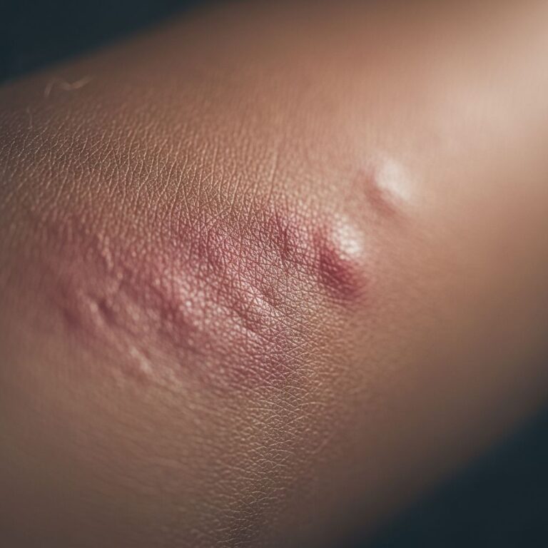
The
classic triad
defines glomus tumours: severepain
,point tenderness
, andcold sensitivity
(exquisite intolerance to cold exacerbates symptoms).- Appearance: Small (1–7 mm), firm, reddish-purple or bluish nodule, often subungual causing nail ridging/deformity.
- Pain characteristics: Paroxysmal, lancinating, worse at night or with pressure/temperature change. Relieved by tourniquet (Hildreth sign).
- Location: Periungual (fingernails > toenails), fingertip pulp, palm. Rare sites: ear, nose.
Multiple lesions present as clustered nodules or plaque-like vascular malformations.
Diagnosis of glomus tumour
Clinical suspicion arises from history and triad; confirmation requires imaging and/or biopsy.
Clinical tests
- Love test: Pinprick pressure elicits severe pain (100% sensitive).
- Hildreth test: Tourniquet reduces pain/tenderness (92% sensitive, 91% specific).
Imaging
**MRI** is gold standard: early T1 isointense, T2 hyperintense ‘nodule in a snail shell’ with punctate enhancement. US shows hypoechoic mass with hypervascularity on Doppler. X-ray may reveal phalangeal erosion in chronic cases.
Histology
Biopsy reveals uniform glomus cells (round/oval nuclei, eosinophilic cytoplasm) in sheets/clusters around capillary channels, branching pattern. No atypia/mitoses.
Glomus tumour pathology
Glomus tumours are dermal, well-demarcated, comprising vascular cores rimmed by glomus cells (bland nuclei, sharp borders). Three patterns exist:
| Type | Features |
|---|---|
| Solid glomus tumour | Predominant glomus cells > vascularity |
| Glomangioma | Dilated vascular channels lined by glomus cell layer |
| Glomangiomyoma | Added smooth muscle/spindled cells |
Immunohistochemistry: Glomus cells +ve for SMA, muscle-specific actin; vessels +ve for CD34/CD31. Negative: cytokeratins, S100.
Differential diagnosis
- Eccrine spiradenoma: Dual cell population, ductal differentiation, cytokeratin +ve.
- Intradermal naevus: S100/Melan-A +ve.
- Angioma: Lacks glomus cells.
Types of glomus tumour
- Solitary glomus tumour: Classic painful subungual nodule.
- Multiple glomus tumours: Familial, less painful, acral.
- Glomuvenous malformation (glomangioma): Congenital, plaque-like, GLMN mutation.
- Malignant glomus tumour: Rare (<1%), larger, atypical, metastatic potential.
Treatment of glomus tumour
**Surgical excision** is curative for solitary lesions. Outpatient procedure: nail avulsion, precise tumour shelling under loupe magnification, layered closure. Immediate pain relief; nail regrows normally in 3–4 months.
- Recurrence: 5–20% if incomplete excision.
- Multiple lesions: Selective excision or sclerotherapy/laser for symptomatic sites.
- Malignant: Wide excision ± lymph node dissection.
Conservative options (NSAIDs, sclerotherapy) offer temporary relief but high recurrence.
Complications of glomus tumour
- Nail dystrophy/ anonychia from chronic pressure.
- Phalangeal erosion.
- Recurrence (1/5 cases).
- Rare malignancy with metastasis.
Prevention of glomus tumour
No prevention exists for sporadic tumours. Genetic counselling for familial multiple glomus tumour families.
Related topics
- Glomuvenous malformations
- Subungual exostosis
- Eccrine spiradenoma
- Onychomatricoma
Frequently Asked Questions
Q: How painful is a glomus tumour?
A: Extremely painful—one of the most painful benign skin lesions, with paroxysmal attacks triggered by touch, cold, or pressure.
Q: Can glomus tumours be cancerous?
A: <1% are malignant (glomangiosarcoma), characterised by size >1 cm, atypia, mitoses, and deep location.
Q: Is MRI always needed for diagnosis?
A: MRI confirms 90–100% but clinical triad + Love test often suffices for excision without preoperative imaging.
Q: What if I have multiple glomus tumours?
A: Suggests inherited glomuvenous malformation; genetic testing recommended. Treat only symptomatic lesions.
Q: Does the tumour come back after surgery?
A: 5–20% recurrence if margins positive; complete excision under magnification minimises risk.
References
- Glomus Tumor — The Arm Doc. Accessed 2026. https://thearmdoc.co.uk/glomus-tumor/
- Glomus tumour pathology — DermNet NZ. Accessed 2026. https://dermnetnz.org/topics/glomus-tumour-pathology
- Glomus tumours — DermNet NZ. 2008. https://dermnetnz.org/topics/glomus-tumour
- Glomus tumor under the microscope (pathology) — YouTube (KiKo Healthcare). Accessed 2026. https://www.youtube.com/watch?v=Hfe4cAr2n60
- Atlas of Head and Neck Pathology: Glomus Tumor — Ohio State University Medicine. Accessed 2026. https://medicine.osu.edu/-/media/files/medicine/departments/otolaryngology/atlas-of-head-and-neck-pathology/g/glomustumor2.pdf
Similar Articles


Read full bio of Sneha Tete