Granular Cell Tumours: Benign Soft Tissue Neoplasms
Comprehensive guide to granular cell tumours: diagnosis, clinical features, and treatment options.

Introduction
Granular cell tumours are rare, generally benign soft tissue neoplasms believed to originate from Schwann cells, which are cells that provide myelin insulation to nerves. These tumours can occur in various anatomical locations including the skin, tongue, breast, gastrointestinal tract, and respiratory tract. Also known as granular cell myoblastoma or Abrikossoff tumour, these lesions display characteristic pathologic findings when examined under a microscope.
The vast majority of granular cell tumours are benign lesions, with less than 2% demonstrating malignant behaviour. While clinically and radiologically they can sometimes mimic malignancy due to their infiltrative growth patterns and poorly defined margins, proper histological evaluation and immunohistochemical analysis can distinguish benign from malignant variants.
Demographics and Epidemiology
Granular cell tumours are rare neoplasms that can occur in anyone, though they display specific demographic patterns. These tumours tend to be more common in females and dark-skinned people. The majority of cases develop between the fourth to sixth decades of life, though the condition can present at any age.
Approximately one-third of all granular cell tumours occur in the skin, with the remaining cases found on the tongue or internal organs. When cutaneous involvement occurs, they most frequently appear on the head and neck region. Multiple granular cell tumours may occur in up to 25% of cases, and multiple lesions are more common in dark-skinned populations.
Causes and Genetic Factors
The exact etiology of granular cell tumours remains incompletely understood. Certain genetic mutations have been linked to these neoplasms, although the precise mechanisms of tumorigenesis remain poorly characterized. In some case reports, multiple granular cell tumours have been associated with certain rare syndromes, including:
- Leopard syndrome
- Noonan syndrome
- Neurofibromatosis type I
These associations suggest a potential genetic predisposition in select patient populations, though such syndromic presentations remain uncommon.
Clinical Features and Presentation
Granular cell tumours present as firm, painless, well-defined nodules that are typically hard upon palpation. The clinical appearance commonly manifests as a red or brown nodule, though lesion colour can vary considerably. Described colours include white, grey, brown, or tan, particularly in dark-skinned individuals where colour variation is more frequently observed.
These tumours occur most commonly in the skin or subcutaneous tissue in 30–40% of cases. When subcutaneous involvement occurs, the nodule typically presents as a small, hard, painless lesion in the dermis or subcutis. The tumour usually remains solitary, though multiple presentations occur in approximately 25% of cases.
Clinical features of granular cell tumours include:
- Firm, painless nodule
- Well-defined borders (though histologically margins may be poorly defined)
- Hard consistency upon palpation
- Variable colour presentation (red, brown, white, grey, or tan)
- Slow growth pattern
- Predilection for head and neck region
Features Suggesting Malignancy
While most granular cell tumours are benign, certain clinical and histopathological features may suggest malignant potential. Clinical features that may indicate malignancy include characteristics such as rapid growth, larger size, and specific histological patterns. Pathological predictors of malignancy include:
- Large tumour size greater than 5 cm
- Deep soft tissue location
- Tumour cell necrosis
- Increased cellularity
- Greater than 2 mitotic figures per 10 high power fields
- Vascular invasion and metastasis
However, it is important to note that none of these findings are pathognomonic of malignancy, and vascular invasion with metastasis may be the only definitive indicator of malignant tumours. Metastatic granular cell tumours involve less than 2% of all cases.
Histological Classification System
A comprehensive classification system was established by Fanburg-Smith et al. in 1998, dividing granular cell tumours into three categories based on six histological features:
- Nuclei pleomorphism: Variation in nuclear size and shape
- Tumour cell spindling: Elongated cell morphology
- Vesicular nuclei with prominent nucleoli: Nuclear membrane characteristics
- Increased nuclear-to-cytoplasmic ratio: Proportion of nuclear material
- Necrosis: Cell death within the tumour
- Increased mitotic rate: Greater than 2 mitoses per 10 high power fields
Classification is determined as follows: benign tumours contain none of these features, atypical variants display 1–2 features, and malignant tumours demonstrate 3 or more features. Additionally, mitoses and/or necrosis in combination with a KI-67 proliferation index greater than 10% are associated with malignant behaviour.
Diagnosis and Histopathology
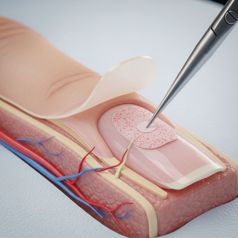
Definitive diagnosis of granular cell tumours is made through skin biopsy. Excision biopsy is the preferred diagnostic approach rather than punch or shave biopsy, as complete histological examination is essential for accurate classification. Excision biopsy also provides the therapeutic benefit of complete tumour removal.
Histological examination reveals characteristic features including:
- Poorly defined tumour composed of nests and ribbons of large polyhedral cells
- Cells separated by thin collagenous bands
- Abundant coarse granular eosinophilic cytoplasm within tumour cells
- Small nuclei with one or two distinct nucleoli
- Absence of cellular atypia in benign cases
The characteristic granules within the cytoplasm of large tumour cells are the hallmark histological finding. Immunohistochemical analysis demonstrates positive reaction for S100 protein and CD68. The proliferation index, assessed by KI-67, is typically less than 1% in benign cases with 0–1 mitotic figures per 10 high power fields.
Radiological Findings
Radiological examination of deep soft tissue granular cell tumours may reveal infiltrative growth patterns that can mimic carcinoma. The poorly defined margins and infiltrative nature of these tumours on imaging may cause initial clinical and radiological examination to suggest a malignant process. This radiological presentation, combined with clinical features, can lead to diagnostic confusion with more aggressive neoplasms. However, the benign nature of most granular cell tumours must be considered in differential diagnosis to avoid unnecessary aggressive interventions.
Differential Diagnoses
Several conditions should be considered in the differential diagnosis of granular cell tumours due to overlapping clinical and histopathological features. Conditions to differentiate include benign and malignant neoplasms with similar presentations. The morphological and immunohistochemical images of granular cell tumours can overlap significantly with both benign and malignant entities, making communication between dermatologists and pathologists essential for accurate diagnosis.
Treatment Approaches
Surgical excision remains the primary treatment modality for granular cell tumours. Treatment approaches differ based on the benign or malignant nature of the lesion:
Benign Cases: Wide surgical excision with appropriate margins is the standard treatment. For benign tumours, a 5 mm margin may be applied, and the majority of cases are cured by simple radical resection. Complete excision provides definitive treatment, and recurrence following adequate initial excision is uncommon.
Malignant Cases: Malignant granular cell tumours require more aggressive intervention including regional lymph node staging and dissection. The effects of chemotherapy and radiotherapy in treating malignant granular cell tumours remain questionable, and surgical resection with appropriate nodal assessment remains the cornerstone of management.
Incomplete initial excision may result in tumour recurrence, emphasizing the importance of adequate surgical margins and complete tumour removal.
Prognosis and Outcomes
The prognosis for benign granular cell tumours is excellent following complete surgical excision. Benign cases are fully cured by complete excision, and follow-up is not necessary after adequate treatment.
However, malignant granular cell tumours demonstrate significantly worse prognosis. These tumours commonly recur and may cause fatal metastatic disease. Malignant variants tend to present with recurrence and metastasis within one year after excision, emphasizing the aggressive nature of these uncommon malignant presentations.
The distinction between benign and malignant granular cell tumours is therefore critically important, as it directly impacts treatment strategy and prognosis. Communication between dermatologists and pathologists is essential, as pathology may provide false reassurance by evaluating a benign-appearing part of a clinically malignant tumour.
Clinical Considerations for Dermatologists
Although granular cell tumours are rare clinically, dermatologists should maintain awareness of their existence and characteristic presentation. Several key considerations should guide clinical management:
- Recognition of the typical presentation as a firm, painless nodule on the head and neck
- Understanding that most cases are benign but malignant variants exist
- Awareness of histological and immunohistochemical features that distinguish benign from malignant disease
- Recognition that radiological findings may mimic malignancy despite benign pathology
- Implementation of excision biopsy as the diagnostic standard
- Ensuring adequate surgical margins for optimal outcomes
- Understanding that follow-up is unnecessary after complete benign tumour excision
Frequently Asked Questions
What is the origin of granular cell tumours?
Granular cell tumours are believed to originate from Schwann cells, which are cells that provide myelin insulation to nerves. This neural origin explains their classification as neural tissue neoplasms.
How common are granular cell tumours?
Granular cell tumours are rare soft tissue neoplasms. Approximately one-third occur in the skin, with the remainder found on the tongue or internal organs. They are more common in females and dark-skinned populations, typically presenting in the fourth to sixth decades of life.
Can granular cell tumours be malignant?
Yes, although less than 2% of granular cell tumours are malignant, malignant variants do exist. Malignant tumours commonly recur and may cause fatal metastatic disease within one year after excision, making accurate diagnosis essential.
What is the best diagnostic approach?
Excision biopsy is the preferred diagnostic method rather than punch or shave biopsy. This approach allows for complete histological examination and simultaneous therapeutic removal of the lesion.
Is follow-up necessary after treatment?
Follow-up is not necessary after complete excision of benign granular cell tumours. However, malignant cases require appropriate staging, surveillance, and management due to their potential for recurrence and metastasis.
What immunohistochemical markers are positive in granular cell tumours?
Granular cell tumours typically show positive immunohistochemical reactions for S100 protein and CD68. The KI-67 proliferation index is generally less than 1% in benign cases.
References
- Granular cell tumour of the skin — Pakistan Journal of Rehabilitation and Restorative Surgery (PRRS). 2023. https://www.prrsjournal.com/article/view/189
- An update on cutaneous granular cell tumours for dermatologists — PubMed/NCBI. 2022. https://pubmed.ncbi.nlm.nih.gov/35727729/
- Neural Tissue Abnormalities: Granular Cell Tumor — Perri Dermatology. https://perridermatology.com/dr-perris-blog/neural-tissue-abnormalities-granular-cell-tumor/
- Granular Cell Tumours — DermNet NZ. 2023. https://dermnetnz.org/topics/granular-cell-tumour
Similar Articles


Read full bio of Sneha Tete