Granuloma Faciale: Diagnosis, Treatment, And Outlook
Rare chronic facial skin condition: causes, diagnosis, treatment options and management strategies.

Granuloma faciale is a rare, benign, chronic inflammatory dermatosis primarily affecting the face, characterised by reddish-brown to violaceous plaques or nodules. It typically presents as single or multiple asymptomatic lesions that persist for months to years, often resistant to initial therapies but manageable with targeted treatments.
What is granuloma faciale?
Granuloma faciale (GF), first described in 1945 as an eosinophilic granuloma of the skin, is an uncommon condition with distinctive clinical and histological features. It manifests as firm, reddish-brown or violaceous plaques, often with a peau d’orange appearance due to follicular prominence and superficial telangiectasias. Lesions are predominantly facial, especially on the cheeks, nose, forehead, or periorbital areas, but extrafacial involvement on the trunk, extremities, or nasal mucosa occurs rarely.
The condition follows a chronic course with recurrent acute phases, showing both acute inflammation and chronic fibrosis histologically. GF is not associated with systemic disease, and its appearance is mainly cosmetic, though some patients report mild tenderness, pruritus, or stinging.
Who gets granuloma faciale?
Granuloma faciale predominantly affects adults aged 40–60 years, with a marked male predominance (male-to-female ratio approximately 3:1). It is rare in children and equally uncommon across ethnic groups, though lighter skin types may highlight lesions more visibly.
- Peak incidence: Middle-aged adults.
- Gender: More common in males.
- Risk factors: Possible associations with sun exposure, trauma, or hypersensitivity, but unproven.
What causes granuloma faciale?
The exact aetiology of granuloma faciale remains unknown. Proposed mechanisms include:
- Immune-mediated vasculitis: Leukocytoclastic vasculitis with perivascular inflammation, eosinophils, and occasional immune complex deposits (IgG, IgA, IgM, C3, C1q).
- Cytokine dysregulation: Interferon-γ (IFN-γ) production by CD4+ T cells driving chronic inflammation.
- Triggers: Hypersensitivity reactions, infections, trauma, actinic exposure, or radiation therapy.
- No links to systemic autoimmune diseases or internal malignancies.
Vascular changes, such as concentric fibrosis around vessels and red cell extravasation (causing brown haemosiderin pigmentation), are hallmarks.
What are the clinical features of granuloma faciale?
Patients typically notice a solitary spot or lesion on the face that enlarges or multiplies over weeks to months. Key features include:
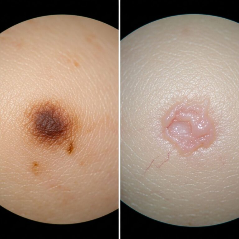
- Appearance: Reddish-brown to violaceous, smooth plaques or nodules (0.5–2 cm), often with telangiectasias, follicular accentuation, and hyperkeratosis.
- Symptoms: Usually asymptomatic; occasional itch, tenderness, or cosmetic distress.
- Distribution: Central face (cheeks, nose, forehead); rare extrafacial sites.
- Progression: Slow growth, chronic persistence; spontaneous resolution uncommon.
Dermoscopy reveals linear vessels, globules, and white scaling, aiding differentiation from sarcoidosis or lupus.
Diagnosis
Diagnosis relies on clinical suspicion confirmed by skin biopsy, essential to exclude mimics.
Histology
Characteristic findings:
- Dermal infiltrate of lymphocytes, eosinophils, neutrophils; mixed acute/chronic features.
- Grenz zone: Sparing of papillary dermis.
- Leukocytoclastic vasculitis: Endothelial swelling, fibrinoid necrosis (mild).
- Haemosiderin deposits: Brown pigmentation.
- Perivascular fibrosis in chronic lesions.
Laboratory tests (e.g., ANA, VDRL, eosinophil count) are normal except occasional mild eosinophilia.
Differential diagnosis
| Condition | Key Distinguishing Features |
|---|---|
| Sarcoidosis | Non-caseating granulomas; systemic involvement; apple-jelly nodules on diascopy. |
| Discoid lupus erythematosus | Scarring, atrophy, photosensitivity; interface dermatitis on biopsy. |
| Erythema elevatum diutinum | Extremity lesions; more neutrophilic; IgA deposits. |
| Lymphocytoma cutis | Lymphocytic nodules; B-cell predominant. |
| Angiolymphoid hyperplasia with eosinophilia | Vascular proliferation; younger patients. |
Biopsy resolves most differentials.
What is the treatment for granuloma faciale?
GF is notoriously treatment-resistant, with no randomised trials due to rarity; evidence from case series and reports. Start with topicals; escalate as needed. Spontaneous remission rare.
First-line: Topical therapies
- Tacrolimus 0.1% ointment BID: Most successful; calcineurin inhibitor blocks T-cell activation. Clearance in many cases after 4–12 weeks.
- High-potency topical corticosteroids: E.g., clobetasol; initial control but relapse common.
- Topical dapsone gel: Anti-inflammatory; promising in small series.
- Pimecrolimus 1% cream: Alternative calcineurin inhibitor.
Second-line: Intralesional/systemic
- Intralesional corticosteroids: Triamcinolone 10–20 mg/mL; repeated injections for thick plaques.
- Oral dapsone 50–100 mg/day: Partial response; monitor for haemolysis.
- Systemic corticosteroids: Short courses; not for long-term.
Advanced therapies
- Laser (PDL, CO2, Nd:YAG): Pulsed dye laser (PDL) effective with purpura induction; 2–3 sessions.
- Cryotherapy: Liquid nitrogen; for small lesions.
- Surgery/dermabrasion: Excision or ablation for refractory cases; scarring risk.
- Others: Hydroxychloroquine, clofazimine, PUVA, TNF-α inhibitors (e.g., adalimumab).
Treatment algorithm: Topicals (tacrolimus/dapsone) → Intralesional steroids → Lasers/cryo → Systemic.
Prevention
No proven prevention; sun protection may help as actinic exposure is implicated. Regular follow-up for recurrence.
Outlook
Lesions persist chronically but are benign; treatments achieve remission in 60–80% with topicals/lasers, though relapse occurs in 20–30%. Cosmetic improvement enhances quality of life.
Frequently Asked Questions (FAQs)
Is granuloma faciale dangerous?
No, it is benign and not linked to cancer or systemic disease.
Does granuloma faciale go away on its own?
Rarely; most require treatment.
What is the best treatment for granuloma faciale?
Topical tacrolimus is first-line based on case series.
Can granuloma faciale appear outside the face?
Yes, rarely on trunk or limbs (extrafacial GF).
How long does treatment take?
Weeks to months; maintenance may be needed.
References
- Granuloma Faciale Treatment: A Systematic Review — Department of Dermatology and Allergology, University Hospital Jena. 2021-03-18. https://www.medicaljournals.se/acta/content/html/10.2340/00015555-2784
- Granuloma faciale: a rare disease from a dermoscopy perspective — PMC/NCBI. 2013-12. https://pmc.ncbi.nlm.nih.gov/articles/PMC3875968/
- Granuloma Faciale – MD Searchlight — MD Searchlight. 2024. https://mdsearchlight.com/skin-problems-and-treatments/granuloma-faciale/
- Granuloma faciale – DermNet — DermNet NZ. 2023. https://dermnetnz.org/topics/granuloma-faciale
- Granuloma Faciale – StatPearls — NCBI Bookshelf. 2023-07-17. https://www.ncbi.nlm.nih.gov/books/NBK539832/
- Granuloma faciale – Primary Care Dermatology Society — PCDS. 2024. https://www.pcds.org.uk/clinical-guidance/granuloma-faciale
Similar Articles


Read full bio of medha deb