Granuloma: 6 Skin Conditions, Diagnosis & Treatment Guide
Comprehensive guide to granulomas: chronic inflammatory skin patterns, types, causes, diagnosis, and management strategies.

A
granuloma
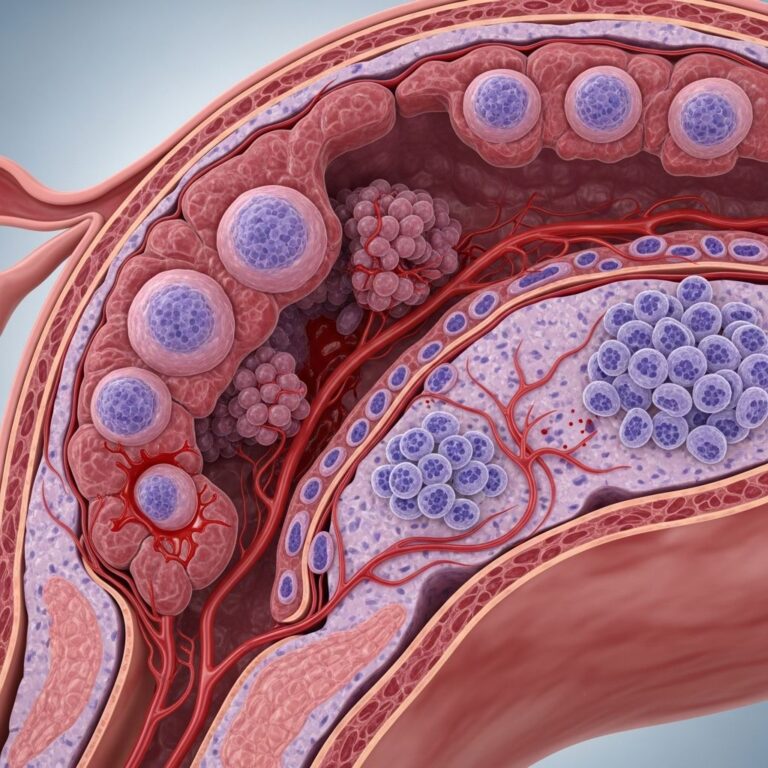
is a specific chronic inflammatory pattern observed in skin biopsies, characterized by the localized aggregation ofhistiocytes
—specialized immune cells derived from monocytes—often accompanied by other inflammatory cells such as plasma cells, eosinophils, or neutrophils. This pattern may include areas of necrosis (tissue death), vasculitis (inflammation of blood vessels), calcification, or foreign bodies, distinguishing it from acute inflammation. Granulomas form as the body’s immune response to persistent stimuli that cannot be easily cleared, such as infections, autoimmune reactions, or non-degradable materials. In dermatology, identifying granulomatous inflammation is crucial for diagnosing a wide array of skin disorders, guiding targeted therapies, and ruling out serious underlying conditions like tuberculosis or malignancy.The formation of granulomas involves a complex interplay of immune cells. Macrophages (histiocytes) aggregate at the site of irritation, attempting to wall off and isolate the offending agent. If unsuccessful, they fuse into multinucleated giant cells, recruiting additional lymphocytes, fibroblasts, and other effectors. This organized structure contrasts with diffuse inflammation seen in conditions like eczema or acute infections. Histopathology remains the gold standard for confirmation, revealing distinctive patterns that narrow differential diagnoses.
What skin conditions are granulomatous?
Granulomatous skin conditions encompass infectious, idiopathic, reactive, and neoplastic processes. They present clinically as papules, nodules, plaques, or ulcers, often with varied colors from red to brown due to hemosiderin deposition. The dermatopathological features vary, but all share the core granuloma structure. Below is a detailed overview of major categories and examples, synthesized from established histopathological patterns.
Infectious Granulomas
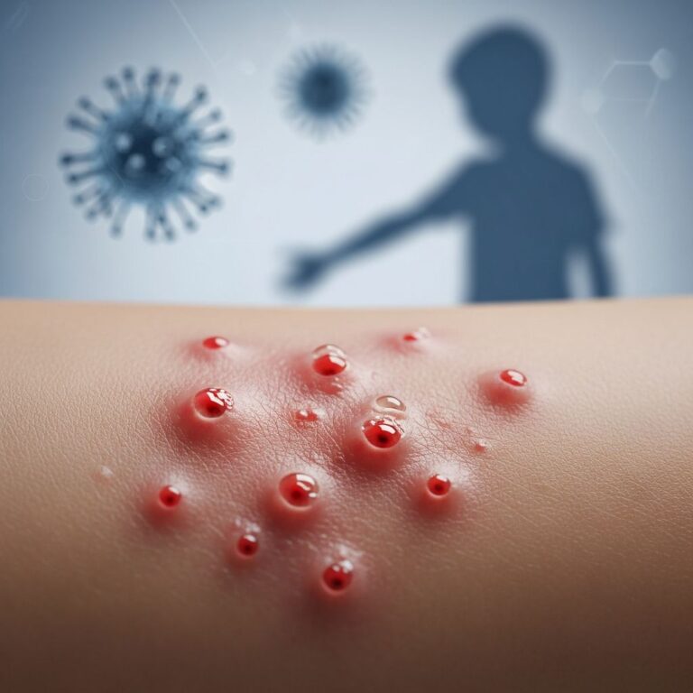
Infections are a leading cause of granulomas, particularly in endemic areas. Mycobacterial infections like tuberculosis produce caseating granulomas with central necrosis resembling cheese (caseation). Leprosy (Mycobacterium leprae) shows foamy histiocytes in non-caseating forms. Fungal infections (e.g., histoplasmosis, cryptococcosis) feature yeast forms within macrophages, identifiable via special stains like PAS or GMS. Parasitic infestations, such as leishmaniasis, elicit granulomas around amastigotes. Bacterial causes include syphilis (gummas) and actinomycosis with sulfur granules. Viral triggers are rarer but include molluscum contagiosum with granulomatous reactions.
- **Tuberculosis**: Caseating granulomas with Langhans giant cells; acid-fast bacilli on Ziehl-Neelsen stain.
- **Leprosy**: Tuberculoid form shows epithelioid granulomas; lepromatous has Virchow cells.
- **Deep Fungal Infections**: Suppurative granulomas with organisms visible on silver stains.
Foreign Body Granulomas
These arise when the immune system encounters indigestible materials, leading to macrophage accumulation and giant cell formation. Common triggers include:
- Silica (silicosis-like skin reactions), suture material, tattoo pigments, cosmetic fillers (e.g., hyaluronic acid, bovine collagen), oils, or metal fragments.
- Clinically, they manifest as red-brown papules, nodules, or plaques, sometimes ulcerated or tender.
- Tattoo granulomas often appear months to years post-procedure, confined to ink sites, presenting as erythematous nodules or lichenoid plaques.
Histology shows refractile foreign material surrounded by histiocytes, with polariscopic examination aiding identification of birefringent particles like talc or starch.
Granuloma Annulare
**Granuloma annulare (GA)** is a benign, inflammatory dermatosis typified by annular plaques of smooth, discolored papules with necrobiotic (collagen degeneration) granulomas on biopsy. It predominantly affects children and young adults (female:male 2:1), though generalized forms occur in older patients.
- **Localized GA**: Most common; rings on dorsal hands, feet, elbows; skin-colored or red, smooth surface with central depression.
- **Generalized GA**: Widespread symmetric papules in large rings on trunk; itchy, associated with diabetes or HIV.
- **Perforating GA**: Yellowish umbilicated papules with crusting; common in Hawaiians or HIV; dermoscopy shows perforations.
- **Atypical GA**: Interstitial forms mimicking other granulomatous dermatitis.
Pathogenesis involves TNF-alpha mediated hypersensitivity to dermal components, triggered by trauma or infections.
Granuloma Faciale
A rare, benign facial disorder with reddish-brown plaques or nodules, often on cheeks. Biopsy reveals dermal eosinophils, leukocytoclastic vasculitis, and a Grenz zone (spared papillary dermis). Hemosiderin imparts brown hue; diagnosis excludes erythema elevatum diutinum or lymphoma.
Granulomatous Dermatitis
This encompasses interstitial and palisading neutrophilic types.
Interstitial granulomatous dermatitis (IGD)
features ‘rope sign’—linear cords on flanks—linked to autoimmune diseases like rheumatoid arthritis or drugs. Histology: lymphocytes, histiocytes in interstitial dermis, collagen degeneration.Palisading neutrophilic granulomatous dermatitis
: Neutrophils rimming necrobiotic areas; associated with systemic vasculitis or rheumatoid nodules.Other Idiopathic and Reactive Granulomas
- **Necrobiosis lipoidica**: Yellow-brown plaques on shins; diabetes-associated; palisading granulomas with atrophied dermis.
- **Cutaneous sarcoidosis**: ‘Apple-jelly’ nodules; naked granulomas without caseation.
- **Rheumatoid nodules**: Subcutaneous; palisading around fibrinoid necrosis.
- **Drug-induced**: Interstitial patterns from TNF inhibitors or others.
Diagnosis of Granulomatous Conditions
Clinical suspicion arises from persistent nodules or plaques unresponsive to standard therapies.
Skin biopsy
is definitive, with processing including H&E stains, special stains (e.g., Ziehl-Neelsen for AFB, GMS for fungi), immunohistochemistry (CD68 for histiocytes), and polarization for foreign bodies. Clinicopathological correlation is essential; for example, annular hand lesions suggest GA, while facial plaques point to granuloma faciale. Additional tests include cultures, PCR for infections, or blood work for systemic associations like ANCA in vasculitis. Dermoscopy aids in perforating GA or tattoos.| Condition | Key Histology | Common Sites | Triggers/Associations |
|---|---|---|---|
| Granuloma Annulare | Necrobiotic collagen, palisading histiocytes | Hands, feet | Idiopathic, trauma, HIV |
| Foreign Body Granuloma | Multinucleated giants around foreign material | Tattoos, injection sites | Fillers, silica, ink |
| Granuloma Faciale | Eosinophils, vasculitis, Grenz zone | Face | Unknown |
| Interstitial Granulomatous Dermatitis | Interstitial histiocytes, fragmented collagen | Trunk flanks | Autoimmune drugs |
Treatment Approaches
Treatment targets the underlying cause where possible. Infectious granulomas require antimicrobials (e.g., rifampin for TB). Foreign body reactions may resolve spontaneously or need excision/laser (Q-switched for tattoos). Idiopathic forms like GA often self-resolve; options include topical/intralesional steroids, cryotherapy, phototherapy, or systemic isotretinoin/NSAIDs for generalized cases. Granuloma faciale responds to tacrolimus or dapsone. Granulomatous dermatitis may remit with antimalarials or biologics like etanercept. Monitoring for recurrence or systemic links is advised.
Frequently Asked Questions (FAQs)
What causes a granuloma on the skin?
Granulomas result from immune responses to infections (e.g., TB, fungi), foreign materials (tattoos, fillers), or idiopathic reactions like granuloma annulare.
Do granulomas go away on their own?
Many, like localized GA or collagen filler reactions, resolve in months to years; others persist or recur without treatment.
Is a granuloma cancerous?
No, granulomas are inflammatory; however, biopsy rules out mimics like cutaneous lymphoma or metastases.
How is granuloma annulare treated?
Often observation suffices; steroids, cryotherapy, or UV therapy for persistent lesions.
Can tattoos cause granulomas?
Yes, tattoo ink triggers foreign body granulomas, treatable with lasers or steroids.
This article provides an overview; consult a dermatologist for personalized diagnosis and management. Granulomas highlight the skin’s role as an immune sentinel, with biopsy unlocking therapeutic paths.
References
- Granuloma faciale — DermNet NZ. 2023. https://dermnetnz.org/topics/granuloma-faciale
- Granuloma annulare — DermNet NZ. Reviewed 2025. https://dermnetnz.org/topics/granuloma-annulare
- Foreign body granuloma — DermNet NZ. 2023. https://dermnetnz.org/topics/foreign-body-granuloma
- Granulomatous dermatitis — DermNet NZ. 2023. https://dermnetnz.org/topics/granulomatous-dermatitis
- Granuloma — DermNet NZ. 2023. https://dermnetnz.org/topics/granuloma
- Granulomatous Disorders of the Skin — Wiley Online Library (Bourke). 2014. https://onlinelibrary.wiley.com/doi/abs/10.1002/9781118441213.rtd0098
Similar Articles


Read full bio of medha deb