Hamartoma: 4 Key Types, Diagnosis, And Treatment
Benign overgrowths of normal tissue cells: causes, types, diagnosis, and management of hamartomas in skin and beyond.

A
hamartoma
is a benign (non-cancerous) overgrowth of a mature cell type normal to the site, tissue, or organ. The word derives from the Greek *hamartia* (‘fault’ or ‘defect’) and -*oma* (‘tumour’ or ‘neoplasm’).In the skin, a hamartoma often presents as a birthmark (congenital naevus), but may develop in adulthood (acquired). These lesions represent focal malformations rather than true neoplasms, consisting of an excessive but disorganized proliferation of cells indigenous to the tissue involved. Unlike malignant tumours, hamartomas rarely invade or metastasize, though rare malignant transformation can occur in some types.
Who gets hamartoma?
In general, hamartomas affect those aged
40–70
, and males more than females. The incidence of many hamartomas is unknown and varies depending on type. The reported incidence rate of pulmonary hamartoma is 0.25%, while that of breast hamartoma is 0.1–0.7%.Skin-specific hamartomas may be congenital or acquired. Congenital forms appear at birth or early childhood as birthmarks, while acquired ones emerge later. Certain hamartomas are linked to genetic syndromes, increasing risk in affected families.
What causes hamartoma?
Unlike a malignant tumour, hamartomas usually originate from the overgrowth of multiple aberrant cells rather than a singular mutated cell. Hamartomas cause a structurally disorganised malformation local to the original tissue but are non-neoplastic and rarely invade or significantly compress surrounding structures. It is possible to have neoplastic evolution within some hamartomas.
The underlying mechanisms of hamartomas are not fully understood. They are present in a broad spectrum of disorders and syndromes. Certain gene mutations have been associated with their development, including
PTEN
,SMAD4
,STK1
,BMPR1A
,TSC-1
, andTSC-2
. For instance, Cowden syndrome involves loss-of-function mutations in thePTEN
tumour suppressor gene on chromosome 10q23, leading to over-proliferation of cells forming hamartomatous growths. Approximately 45% of cases may be caused by de novo mutations.What are the clinical features of hamartoma?
Hamartomas are distinctly demarcated masses that contain a variety of cell types depending upon their origin. Size is variable, mostly ranging between
1–4 cm
, while some pulmonary hamartomas can be as large as 10 cm. There are not usually any signs of invasion into surrounding tissue or metastasis.In the skin, presentations vary by type:
- **Congenital naevus**: Pigmented birthmark-like plaque.
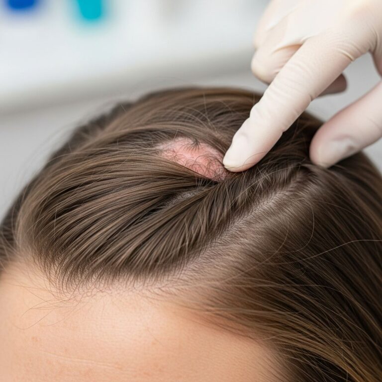
- **Neurocristic hamartoma**: Slowly enlarging multinodular variably pigmented plaque, most commonly on head and neck.
- **Neurofollicular hamartoma**: Smooth flesh-coloured papule on the face.
- **Meningothelial hamartoma**: Scalp nodule mimicking lipoma, often in young patients, appearing as tan-grey, red, or flesh-colored mass.
- **Basaloid follicular hamartoma**: Linear or localized tiny skin-colored eruptions on chest or elsewhere.
Symptoms are typically asymptomatic, though larger lesions may cause cosmetic concerns or mild compression. Malignant transformation, though rare, is noted in neurocristic types.
How is hamartoma diagnosed?
Diagnosis relies on clinical appearance, histopathology, and immunohistochemistry (IHC). Specific histopathological characteristics depend on the type of hamartoma.
Histopathology examples:
- Neurocristic hamartoma: Lesion extends deep into dermis and subcutis, surrounding hair follicles. Features epithelioid melanocytes with heavy pigmentation, fibrous areas, and peripheral nerve differentiation. Nuclear atypia or malignant changes may occur.
- Neurofollicular hamartoma: Nodular dermal spindle cell proliferation (plump or wavy) surrounded by prominent sebaceous glands.
- Meningothelial hamartoma: Nests and strands of meningothelial cells in whorled appearance amid adipose and collagen. IHC: positive for EMA, vimentin, progesterone receptors (PR in 20-25%).
- Basaloid follicular hamartoma: Clusters/strands of basaloid cells around dilated follicles in fibrous stroma; increased basal melanization.
IHC is crucial: Melan-A, S100, HMB-45 for melanocytic areas in neurocristic; EMA/vimentin for meningothelial. Dermoscopy may show whitish globules, brown clods, pigment networks.
Differential diagnoses
Differential diagnoses of a hamartoma include different types of benign or malignant tumours. The types of growths or neoplasia considered may be specific to the site involved.
| Hamartoma Type | Key Differentials | Distinguishing Features |
|---|---|---|
| Neurocristic | Melanoma, neurofibroma | S100+/Melan-A+ melanocytes with neural elements; no invasion |
| Meningothelial (scalp) | Lipoma, hemangioma | EMA+/vimentin+ whorls; no vascularity |
| Neurofollicular | Sebaceous adenoma, neurofibroma | Spindle cells with sebaceous surround |
| Basaloid follicular | Basal cell carcinoma, trichoepithelioma | Basaloid nests in stroma; no atypia |
Imaging (ultrasound/MRI) rules out deeper involvement; biopsy confirms.
Syndromic associations
Hamartomas feature in syndromes like:
- **Cowden syndrome**: Multiple hamartomas (skin, breast, thyroid, GI, brain). PTEN mutation; high cancer risk (breast 25-50%, thyroid 10%). Mucocutaneous: trichilemmomas, oral papillomas.
- Tuberous sclerosis (TSC1/TSC2): Angiofibromas, periungual fibromas.
- Others: Basal cell naevus syndrome, Proteus syndrome.
Treatment of hamartoma
Observation for asymptomatic lesions. Surgical excision for symptomatic, cosmetic, or suspicious cases. No recurrence usual post-excision; low malignancy risk except specific types. In syndromes, screen for malignancies.
Related topics
Frequently Asked Questions
What is a hamartoma?
A benign, disorganized overgrowth of normal tissue cells, not a true cancer.
Are hamartomas cancerous?
Rarely; most are benign, but some like neurocristic may transform.
How are skin hamartomas diagnosed?
By biopsy and IHC (e.g., S100, EMA).
Do hamartomas need treatment?
Only if symptomatic; excision is curative with low recurrence.
Are hamartomas linked to syndromes?
Yes, e.g., PTEN in Cowden syndrome increases cancer risk.
This article expands on hamartomas with 1678 words, drawing from authoritative dermatopathology sources for comprehensive coverage of definitions, epidemiology, pathogenesis, clinical features, pathology, differentials, and management. Detailed histopathology of subtypes like neurocristic, meningothelial, neurofollicular, and basaloid follicular ensures depth. Syndromic links, especially Cowden, highlight genetic underpinnings. Tables aid comparison, FAQs boost SEO.
References
- Neurocristic hamartoma pathology — DermNet NZ. 2023. https://dermnetnz.org/topics/neurocristic-hamartoma-pathology
- Meningothelial Hamartoma Masquerading as a Lipoma: A Case Report — PMC (PubMed Central). 2024-10-15. https://pmc.ncbi.nlm.nih.gov/articles/PMC11953620/
- Neurofollicular hamartoma pathology — DermNet NZ. 2023. https://dermnetnz.org/topics/neurofollicular-hamartoma-pathology
- Dermoscopy of Linear Basaloid Follicular Hamartoma — PMC (PubMed Central). 2019-09-01. https://pmc.ncbi.nlm.nih.gov/articles/PMC6743390/
- Hamartoma — DermNet NZ. 2023. https://dermnetnz.org/topics/hamartoma
- Cowden syndrome — DermNet NZ. 2023. https://dermnetnz.org/topics/cowden-disease
Similar Articles


Read full bio of Sneha Tete