Heart’s Electrical System: Anatomy and Function
Understanding how electrical impulses control your heart's rhythm and coordinate its pumping action.

Understanding the Heart’s Electrical System
The heart is more than just a muscular pump—it is an intricate organ controlled by a sophisticated electrical system that coordinates the contraction of its four chambers and regulates heart rate. This electrical conduction system ensures that blood is pumped efficiently throughout your body, delivering oxygen and nutrients to every tissue and organ. Without this carefully orchestrated system, the heart would not be able to maintain the precise rhythm necessary to sustain life.
The purpose of the heart’s electrical system is twofold: to coordinate the pumping of the four chambers and to control the heart rate so that it speeds up and slows down in response to the body’s changing demands. When you exercise, your heart rate accelerates to deliver more oxygen-rich blood to your muscles. When you rest, it slows down. This remarkable adaptation is made possible by the electrical signals that originate from specialized tissue within the heart.
The Components of the Cardiac Conduction System
The heart’s electrical system comprises several key structures, each with a specific role in maintaining proper heart rhythm and function. Understanding these components is essential to grasping how your heart works.
The Sinoatrial Node: The Heart’s Natural Pacemaker
The journey of each heartbeat begins in the sinoatrial node, commonly referred to as the SA node. This small mass of specialized electrical tissue is located in the right upper chamber of the heart, known as the right atrium, near the entrance of the superior vena cava. The SA node acts as the heart’s “natural pacemaker,” generating the electrical impulses that initiate every heartbeat.
In a healthy adult at rest, the SA node creates an electrical signal 60 to 100 times per minute. This regular rhythm is what we experience as our normal resting heart rate. The SA node’s ability to generate these impulses automatically makes it unique—it does not require input from the brain to function, though the nervous system can modulate its rate based on bodily needs.
The Atria and Initial Electrical Spread
Once the SA node generates an electrical impulse, this signal spreads rapidly across the cells of both the right and left atria. This electrical activity causes the atrial muscle to contract. The contraction of the atria serves an important purpose: it pushes blood from these upper chambers into the lower chambers, or ventricles, ensuring efficient filling before ventricular contraction occurs.
The timing of atrial contraction is crucial. The atria must contract and empty their blood into the ventricles just before the ventricles begin their own contraction. This sequential timing prevents backup of blood and ensures maximum cardiac output with each heartbeat.
The Atrioventricular Node: The Electrical Gateway
After stimulating the atria, the electrical signal travels downward to the atrioventricular node, commonly called the AV node. The AV node is strategically positioned between the atria and the ventricles and serves as a critical control point in the conduction system. Rather than allowing the electrical impulse to travel instantaneously to the ventricles, the AV node deliberately slows the signal for a very brief period, typically just a fraction of a second.
This seemingly minor delay is actually crucial to heart function. By slowing the electrical impulse at the AV node, the system ensures that the atria have completely emptied their blood into the ventricles before ventricular contraction begins. This allows the ventricles to fill completely, maximizing the volume of blood they can pump with each contraction. Without this deliberate delay, the atria and ventricles would contract nearly simultaneously, and the ventricles would not fill adequately.
The Bundle of His and Bundle Branches
After passing through the AV node, the electrical signal continues its journey down the conduction pathways through a structure called the bundle of His. This bundle of specialized nerve fibers carries the electrical signal from the AV node into the ventricles. The bundle of His is located within the septum—the muscular wall that separates the right and left ventricles.
As the bundle of His extends into the ventricles, it divides into two pathways: the right bundle branch and the left bundle branch. These bundle branches distribute the electrical signal to the right and left ventricles respectively, ensuring that both ventricles receive electrical stimulation at approximately the same time. This synchronized activation allows both ventricles to contract in a coordinated manner, pumping blood efficiently to the lungs and the rest of the body.
The Purkinje Fibers: The Final Conduction Pathway
The electrical signal’s final destination within the heart is reached through the Purkinje fibers, sometimes referred to as the “thin wires” of the conduction system. These specialized muscle fibers extend from the bundle branches into the muscular walls of both ventricles. The Purkinje fibers spread the electrical signal throughout the ventricular muscle, ensuring that all areas of the ventricles receive the stimulation needed to contract uniformly and powerfully.
This comprehensive distribution of electrical activity to all parts of the ventricles is essential for effective pumping. When the entire ventricular wall contracts as one coordinated unit, the heart can generate sufficient pressure to pump blood through the lungs and out to the rest of the body.
How a Heartbeat Occurs: The Complete Electrical Sequence
A single heartbeat involves a carefully choreographed sequence of electrical events. Understanding this sequence provides insight into how an estimated 100,000 heartbeats occur each day to keep us alive and healthy.
The sequence begins when the SA node generates an electrical impulse. This impulse rapidly spreads throughout the right and left atria, causing both atria to contract almost simultaneously. As the atria contract, they push blood into the ventricles below. Meanwhile, the electrical signal reaches the AV node, where it slows momentarily. This pause ensures the atria fully empty before the ventricles contract.
Once the signal passes through the AV node, it travels down the bundle of His and into the bundle branches. The electrical current then reaches the Purkinje fibers, which distribute the signal throughout the ventricular muscle. This stimulation causes the ventricles to contract powerfully, pumping blood out into the arteries—the right ventricle sends blood to the lungs, while the left ventricle sends blood to the rest of the body.
As the ventricles contract and pump out blood, the electrical signal gradually fades, and the heart muscle begins to relax. This relaxation phase allows the ventricles to fill with blood again. The entire cycle, from the SA node’s initial impulse to complete ventricular relaxation, constitutes one heartbeat. This cycle then repeats, maintaining the continuous circulation of blood throughout your life.
Heart Rate Regulation and Adaptation
While the SA node generates heartbeats at a baseline rate of 60 to 100 beats per minute in a resting adult, your heart rate is not fixed. The nervous system and various hormones modulate the SA node’s firing rate based on your body’s needs. During exercise, stress, or illness, your heart rate increases. During rest, relaxation, or sleep, your heart rate decreases.
This flexibility is achieved through the autonomic nervous system, which includes the sympathetic and parasympathetic divisions. The sympathetic nervous system speeds up the heart rate when you need more oxygen and blood flow. The parasympathetic nervous system slows the heart rate during periods of rest and recovery. This dynamic regulation ensures that your cardiovascular system always meets your body’s metabolic demands.
The Role of Backup Pacemakers
While the SA node is the primary pacemaker, the heart has built-in redundancy. If the SA node fails to function properly, other parts of the conduction system can take over as backup pacemakers. The AV node can generate electrical impulses at a slower rate, typically 40 to 60 beats per minute. The bundle branches and Purkinje fibers can generate impulses even more slowly, at 20 to 40 beats per minute. This backup system ensures that even if the primary pacemaker fails, the heart can continue to beat, though potentially at a reduced rate. In cases where these backup systems are insufficient, patients may require a artificial pacemaker implant.
Common Electrical System Disorders
When the heart’s electrical system malfunctions, various arrhythmias—abnormal heart rhythms—can develop. These range from benign conditions to life-threatening situations. Common disorders include atrial fibrillation, where the atria quiver irregularly instead of contracting effectively; ventricular fibrillation, a dangerous condition where the ventricles quiver chaotically; and heart block, where electrical signals are delayed or blocked at the AV node. Understanding the electrical system helps clinicians diagnose and treat these conditions effectively.
Diagnostic Tools: The Electrocardiogram
The electrocardiogram, or ECG, is a diagnostic tool that records the electrical activity of the heart. By placing electrodes on the skin, healthcare providers can visualize the heart’s electrical impulses and detect abnormalities in the conduction system. The ECG shows distinctive waves corresponding to atrial depolarization, ventricular depolarization, and repolarization, providing a real-time snapshot of the heart’s electrical function.
Frequently Asked Questions About the Heart’s Electrical System
Q: What happens if the SA node stops working?
A: If the SA node fails, the AV node or other parts of the conduction system can serve as backup pacemakers, though at a slower rate. In severe cases, a permanent pacemaker may be needed to maintain adequate heart rate.
Q: Can stress affect the electrical system of the heart?
A: Yes, stress activates the sympathetic nervous system, which increases the firing rate of the SA node, causing your heart to beat faster. Chronic stress can contribute to arrhythmias and other cardiac problems.
Q: What is a normal resting heart rate?
A: A normal resting heart rate for adults is typically between 60 and 100 beats per minute. However, athletes may have lower resting rates due to cardiovascular conditioning.
Q: How does exercise increase heart rate?
A: During exercise, your muscles demand more oxygen and blood. The sympathetic nervous system responds by increasing the SA node’s firing rate, causing your heart to beat faster and pump more blood.
Q: What role does the AV node play in heart function?
A: The AV node delays the electrical signal by a fraction of a second, allowing the atria to fully contract and empty into the ventricles before the ventricles begin their contraction, ensuring efficient cardiac output.
Q: Can the heart’s electrical system be repaired?
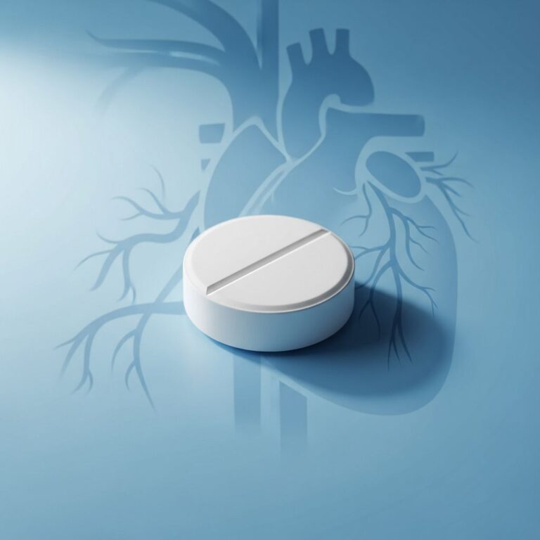
A: Depending on the problem, various treatments exist, including medications, catheter ablation procedures, and artificial pacemakers, which can help restore normal heart rhythm and function.
Maintaining Heart Electrical Health
Keeping your heart’s electrical system healthy involves maintaining overall cardiovascular health. Regular exercise, a balanced diet, stress management, adequate sleep, and avoiding smoking and excessive alcohol consumption all support optimal heart function. Managing conditions like high blood pressure, diabetes, and high cholesterol also protects the electrical system from damage. Regular check-ups with your healthcare provider can help identify and address any issues early.
References
- Electrical System of the Heart — Hobart Cardiology. Accessed December 1, 2025. https://hobartcardiology.com.au/the-normal-heart/electrical-system-heart/
- Anatomy and Function of the Heart’s Electrical System — University of Michigan Health. Accessed December 1, 2025. https://www.ummhealth.org/health-library/anatomy-and-function-of-the-hearts-electrical-system
- Anatomy and Function of the Electrical System — University of Rochester Medical Center. Accessed December 1, 2025. https://www.urmc.rochester.edu/encyclopedia/content?contenttypeid=90&contentid=p01762
- Heart Conduction System (Cardiac Conduction) — Cleveland Clinic. Accessed December 1, 2025. https://my.clevelandclinic.org/health/body/21648-heart-conduction-system
- Our Heart and its Electrical System — Cleveland Clinic Abu Dhabi. Accessed December 1, 2025. https://www.clevelandclinicabudhabi.ae/en/health-byte/heart-and-vascular-health/our-heart-and-its-electrical-system
Similar Articles


Read full bio of medha deb