Hemangioma: 5 Types, Symptoms & Treatment Options
Understanding hemangiomas: benign vascular tumors in infants, their growth phases, symptoms, diagnosis, and effective treatments.

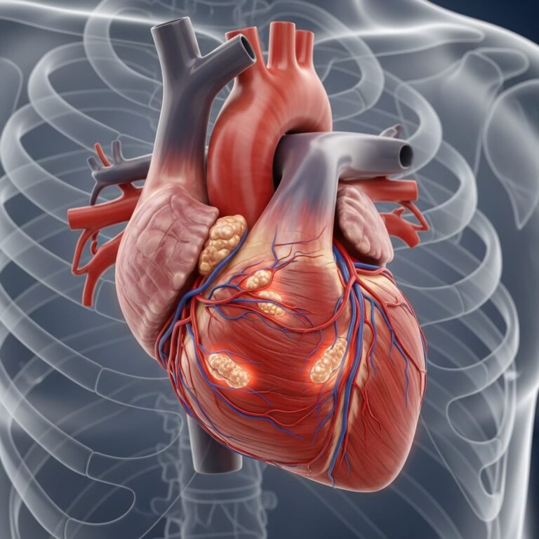
Hemangiomas, often called “strawberry marks,” are the most common benign tumors of infancy, resulting from endothelial cell proliferation in blood vessels. These vascular lesions typically appear in the first weeks of life and follow a predictable pattern of rapid growth followed by gradual involution.
What Is a Hemangioma?
A
hemangioma
is a benign (non-cancerous) tumor composed of extra blood vessels, most frequently occurring in infants. Unlike malignant tumors, hemangiomas do not spread and often resolve spontaneously. They are classified into infantile hemangiomas, which develop after birth, and congenital hemangiomas, present at birth.Infantile hemangiomas (IHs) arise from rapid proliferation of endothelial cells, forming a multinodular pattern supplied by a single arteriole. Histologically, they feature hyperplastic endothelial cells, pericytes, and prominent basement membranes during the proliferative phase, transitioning to dilated vascular lumens and flattened cells in involution.
Congenital hemangiomas differ: rapidly involuting congenital hemangiomas (RICH) shrink within the first year, while non-involuting congenital hemangiomas (NICH) persist.
Symptoms of Hemangioma
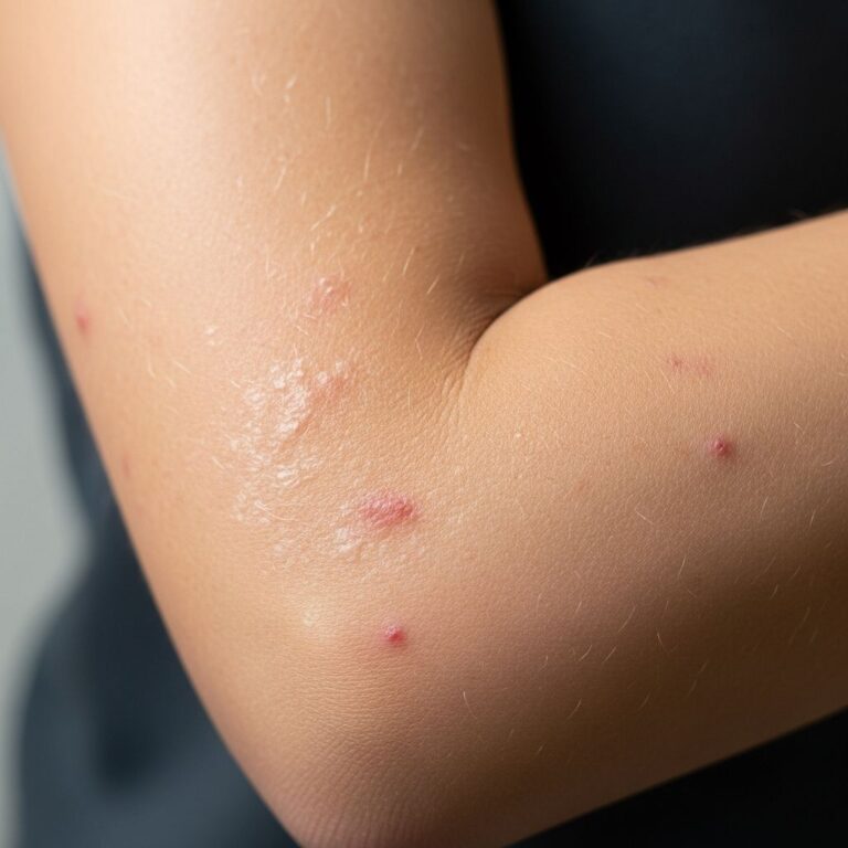
Hemangiomas present as painless, red to blue lesions on the skin, lips, or inside the mouth. They feel soft, warm to the touch, and may be flush with the skin or slightly elevated, sometimes growing from a stalk.
- Superficial hemangiomas: Raised, bright red, lobulated areas in the dermis, resembling strawberries.
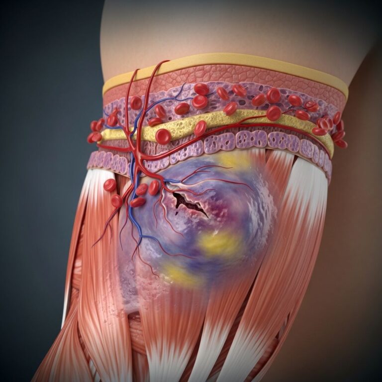
- Deep hemangiomas: Bluish nodules or plaques in the subcutis, appearing subcutaneous.
- Mixed hemangiomas: Combine features of both superficial and deep types.
Superficial lesions may ulcerate or bleed if injured. Deep ones in muscle can cause pain and activity-related swelling; in bones, pain and enlargement. Most are asymptomatic but can lead to complications if in critical areas.
Causes
The exact cause of hemangiomas remains unclear, but they stem from abnormal endothelial cell proliferation forming clusters of small blood vessels (capillaries). Genetic and environmental factors may contribute, though no single trigger is identified.
Infantile hemangiomas are not present at birth but emerge shortly after, linked to placental endothelial cells migrating to the infant’s skin. Risk factors include prematurity, low birth weight, and female sex (girls are affected 4-5 times more than boys).
Diagnosis
Diagnosis is primarily clinical, based on history and physical exam: noting lesion location, size changes, and associated symptoms like pain or swelling.
Key questions include duration, growth patterns, and functional impacts. Palpation assesses texture, depth, and tenderness. High-risk sites (e.g., near eyes, nose, lips) warrant screening for complications.
- Imaging: Ultrasound for deep lesions; MRI if visceral involvement suspected.
- Biopsy: Rarely needed, confirms lobular capillary architecture.
Differentiate from vascular malformations (present at birth, grow proportionally) or other tumors.
Natural History and Growth Phases
Infantile hemangiomas follow a triphasic cycle:
- Proliferative phase (0-12 months): Rapid growth in first 3 months (80% by month 3), slower to 5-8 months; deep ones longer (up to 12 months).
- Plateau phase (6-12 months): Stable size.
- Involution phase (1-10 years): Shrinkage; 50% resolve by age 5, 70% by 7, 90% by 9. Residual changes like fibrofatty tissue, telangiectasia, or lax skin in ~8%.
Congenital types: RICH involute rapidly; NICH do not.
Types of Hemangiomas
| Type | Description | Location | Behavior |
|---|---|---|---|
| Superficial (Capillary) | Bright red, raised, dermal | Head/neck (60%) | Grows then involutes |
| Deep | Bluish subcutaneous nodule | Trunk (25%) | Proliferates longer |
| Mixed | Both superficial/deep features | Extremities (15%) | Variable involution |
| Congenital (RICH) | Fully formed at birth | Various | Rapid involution |
| Congenital (NICH) | Fully formed at birth | Various | Persistent |
Head/neck predominance due to higher vascularity.
When to See a Doctor
Seek care for:
- Functional impairment (vision, breathing, feeding).
- Ulceration, bleeding, or infection.
- Rapid growth or pain.
- Locations: periorbital, nasal tip, lips, anogenital.
Early referral to dermatology or vascular specialists improves outcomes.
Treatment
Most hemangiomas (90%) require no treatment, resolving spontaneously. Interventions target complications, ulceration, or disfigurement.
First-line: Beta-blockers (e.g., oral propranolol 2-3 mg/kg/day). Accelerates involution, reduces size; monitor for bradycardia, hypotension.
- Topical: Timolol for small superficial lesions.
- Corticosteroids: Intralesional for focal; systemic rarely.
- Laser: Pulsed dye for telangiectasia/ulceration; controversial for deep.
- Surgery: For residual fibrofatty tissue, deep muscle/bone involvement, or functional threats. Excision under anesthesia; risks include bleeding, recurrence.
Complications
Ulceration (10%, esp. lips, anogenital). Ophthalmic: amblyopia, astigmatism. Airway obstruction, heart failure in large hepatic. Cosmetic residuals.
Prognosis
Excellent; complete resolution in most by puberty. ~8% need intervention for residuals. Early treatment prevents complications.
Prevention
No known prevention; monitor at-risk infants (preemies). Early screening key.
Frequently Asked Questions (FAQs)
Are hemangiomas cancerous?
No, hemangiomas are benign and do not become cancerous.
Do all hemangiomas go away?
Most infantile ones involute by age 9-10; congenital NICH do not.
Is propranolol safe for babies?
Yes, when monitored; first-line for high-risk cases.
Can hemangiomas cause pain?
Usually not; deep ones in muscle/bone may.
How are hemangiomas diagnosed?
Clinically; imaging/biopsy if needed.
References
- Hemangioma – StatPearls — NCBI Bookshelf. 2023. https://www.ncbi.nlm.nih.gov/books/NBK538232/
- Hemangioma – Symptoms, diagnosis and treatment — BMJ Best Practice. 2023. https://bestpractice.bmj.com/topics/en-us/1041
- Hemangiomas – Vascular Malformations — OrthoInfo – AAOS. 2023. https://orthoinfo.aaos.org/en/diseases–conditions/hemangioma
- Diagnosis and Management of Infantile Hemangioma — American Academy of Pediatrics. 2015-02-01. https://publications.aap.org/pediatrics/article/136/4/786/73896/Diagnosis-and-Management-of-Infantile-Hemangioma
Similar Articles


Read full bio of medha deb