Hemothorax: Causes, Symptoms, Diagnosis & Treatment
Learn about hemothorax: blood in the pleural space, its causes, symptoms, diagnosis methods, and treatment options.
Understanding Hemothorax: A Comprehensive Guide
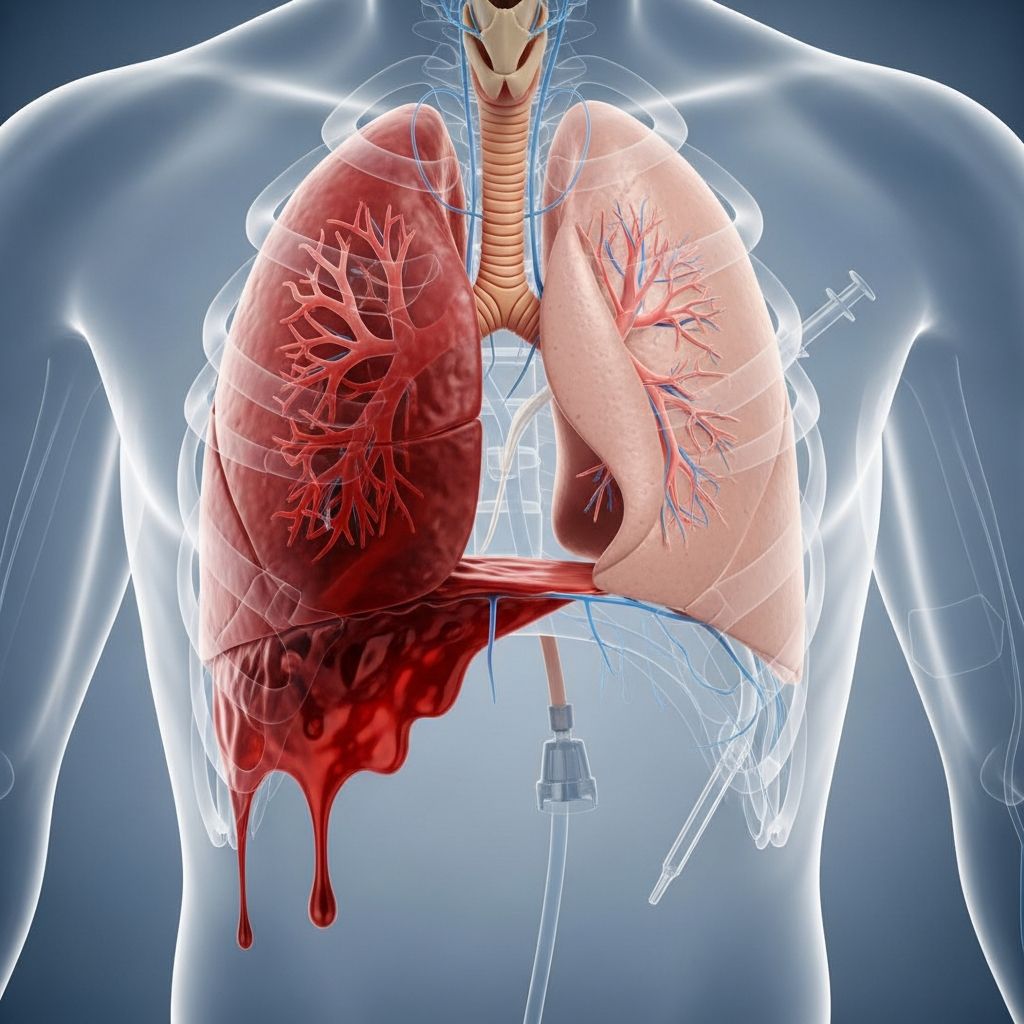
Hemothorax is a serious medical condition in which blood accumulates in the pleural space, the area between your lungs and the inner chest wall. This collection of blood can put significant pressure on your lungs, preventing them from expanding properly and compromising your ability to breathe. Understanding hemothorax is crucial for recognizing symptoms early and seeking immediate medical attention, as this condition requires prompt intervention to prevent life-threatening complications.
What Is Hemothorax?
Hemothorax refers specifically to the accumulation of blood in the pleural cavity, which is the potential space between the visceral pleura (the membrane covering the lungs) and the parietal pleura (the membrane lining the chest wall). Each hemithorax, or side of the chest cavity, can potentially hold up to 40% of a patient’s total circulating blood volume, making this a potentially life-threatening condition when significant bleeding occurs.
The pleural space normally contains only a small amount of fluid that lubricates the surfaces and allows the lungs to move smoothly during breathing. When blood enters this space, it displaces the lung tissue and restricts its expansion, leading to respiratory compromise and potentially severe oxygen deprivation.
Causes of Hemothorax
Hemothorax is most commonly caused by traumatic thoracic injuries. The bleeding can originate from various structures within the chest cavity:
Traumatic Injuries: Hemothorax is a frequent consequence of chest trauma, which can occur from motor vehicle accidents, falls from heights, sports injuries, or penetrating wounds such as stab or gunshot injuries.
Internal Bleeding Sources: Bleeding may arise from damage to several structures, including diaphragmatic injuries, mediastinal structures, pulmonary vessels, pleural membrane injuries, chest wall structures, and occasionally abdominal organs that extend into the thoracic cavity. Injury to intercostal vessels, including the internal mammary arteries and pulmonary vessels, frequently leads to significant bleeding requiring invasive management.
Blunt vs. Penetrating Trauma: Both blunt force trauma and penetrating injuries can cause hemothorax, though the underlying injuries may differ in nature and severity.
Risk Factors and Epidemiology
Hemothorax occurs primarily in individuals who have experienced significant chest trauma. Risk factors include participation in high-impact activities, occupations with injury risks, motor vehicle operation, and any circumstance where severe chest injury might occur. The condition is particularly common in trauma centers and emergency departments, affecting both civilian and military populations exposed to traumatic incidents.
Recognizing Symptoms of Hemothorax
The clinical presentation of hemothorax varies depending on the volume of blood accumulated and the speed of accumulation. Symptoms often overlap with those of pneumothorax (air in the pleural space) and other chest injuries.
Respiratory Symptoms:
– Respiratory distress and difficulty breathing
– Rapid breathing (tachypnea)
– Shortness of breath
– Low oxygen saturation (hypoxia)
Chest Symptoms:
– Severe chest pain, especially with breathing
– Chest wall asymmetry (one side appears different from the other)
– Paradoxical chest wall movement (flail chest), where the chest moves inward during inhalation
Physical Examination Findings:
– Decreased or absent breath sounds on the affected side
– Dullness to percussion over the affected area
Related Stories



– Tracheal deviation (the airway shifts to the opposite side)
– Distended neck veins, suggesting pericardial tamponade or tension pneumothorax
– Signs of chest wall trauma including contusions, abrasions, the characteristic “seat belt sign,” penetrating injuries, or ecchymosis (bruising)
Cardiovascular Symptoms:
– Hypotension (low blood pressure)
– Narrow pulse pressure
– Rapid heart rate (tachycardia)
– Signs of shock in severe cases
Other Signs:
– Facial or neck swelling
– Cyanosis (bluish discoloration of skin)
– Crepitus (crackling sensation under the skin)
– Point tenderness in the chest wall
Diagnosis of Hemothorax
Accurate diagnosis of hemothorax requires a combination of clinical examination, imaging studies, and laboratory tests. Healthcare providers follow systematic trauma protocols to identify and assess the severity of the condition.
Physical Examination: A healthcare provider will conduct a thorough physical examination, listening to the lungs with a stethoscope and assessing vital signs including oxygen saturation, pulse, breathing rate, and blood pressure. The examination focuses on identifying signs of chest trauma and respiratory compromise.
Imaging Studies:
– Chest X-rays: Initial imaging that can reveal blood accumulation in the pleural space, lung collapse, and mediastinal shift
– CT Scan: Provides detailed three-dimensional images and helps identify specific injuries to organs, vessels, and other chest structures
– Ultrasound: May be used to assess pleural fluid and guide procedures
Laboratory Tests: The recommended work-up includes:
– Complete Blood Count (CBC) to establish baseline hemoglobin levels
– Comprehensive Metabolic Panel (CMP) including creatinine assessment
– Troponin levels to evaluate for cardiac contusion
– Coagulation profile to assess bleeding and clotting function
– Type and screen for potential blood transfusion
– Serial troponins and electrocardiogram (EKG) if cardiac injury is suspected
– Serum lactate, which serves as a predictor of mortality in trauma patients (levels above 4 mg/dL correlate with higher mortality)
Pleural Fluid Analysis: When fluid is obtained, analysis can help determine the nature of the fluid and rule out infection or other complications.
Treatment and Management of Hemothorax
The treatment approach for hemothorax depends on the volume of blood accumulated, the patient’s stability, and the ongoing rate of bleeding.
Initial Resuscitation: All patients receive initial resuscitation and management according to Advanced Trauma Life Support (ATLS) protocols. This includes establishing two large-bore intravenous lines, placing patients on cardiac and oxygen monitors, obtaining a 12-lead EKG, and providing supplemental oxygen.
Minimal Hemothorax: When the blood collection is minimal (less than 300 milliliters), treatment is typically conservative. The blood usually reabsorbs naturally over several weeks. If the patient is stable with minimal respiratory distress, operative intervention is not required. Management consists of pain control through analgesia and observation with repeated imaging at 4 to 6 hours and 24 hours after injury.
Moderate to Large Hemothorax: For larger collections, a chest tube (thoracostomy) may be inserted through the chest wall between the ribs to drain blood and air from the pleural space. The chest tube is left in place and attached to suction for several days to allow the lung to re-expand.
Breathing Support: Patients may receive supplemental oxygen, non-invasive airway pressure support such as BiPAP, or in severe cases, endotracheal intubation with mechanical ventilation to support breathing.
Surgical Intervention: Urgent anterior thoracotomy (surgical opening of the chest) is indicated when:
– 1,500 milliliters of blood drains through the chest tube in 24 hours
– 300-500 milliliters drain per hour for 2 to 4 consecutive hours after chest tube insertion
– Great vessel or chest wall injury is present
– Pericardial tamponade develops
Thoracotomy allows rapid assessment of intrathoracic injuries and achievement of hemostasis (stopping the bleeding).
Advanced Procedures: Video-assisted thoracoscopy (VATS) has demonstrated significant effectiveness in managing retained hemothorax. This minimally invasive procedure provides clear visualization of the pleural cavity, enables correct chest tube placement for accurate bleeding control, allows removal of retained clots, and facilitates evacuation and decortication of post-traumatic empyemas. VATS also allows evaluation of suspected diaphragmatic injuries and mediastinal injuries while treating persistent air leaks.
Blood Transfusion: Patients who lose significant amounts of blood may require blood transfusions to maintain adequate blood volume and oxygen-carrying capacity.
Infection Prevention: After treatment, healthcare providers prescribe antibiotics to prevent infection and pain relievers to reduce discomfort during healing.
Complications of Hemothorax
Without prompt treatment, hemothorax can lead to serious complications:
– Collapsed Lung (Pneumothorax): Blood and air accumulation can prevent lung inflation, leading to lung collapse and respiratory failure
– Respiratory Failure: Severe compromise of lung function preventing adequate oxygen exchange
– Empyema: Infection of the pleural fluid, which can occur if hemothorax evacuation is improper or incomplete
– Fibrothorax or Trapped Lung: Fibrosis or scarring of the pleural membranes and underlying lung tissue, resulting in permanent lung dysfunction
– Shock: Severe blood loss can lead to hypovolemic shock
– Death: In severe circumstances with massive bleeding or delayed treatment
– Recurrence: Hemothorax can recur, requiring continued monitoring
Prognosis and Recovery
The outlook for hemothorax depends on several factors: the cause of the hemothorax, the amount of blood loss, how quickly treatment is provided, and the severity of associated injuries. In cases of major trauma, prognosis additionally depends on the severity of the injury and the rate of bleeding.
With prompt diagnosis and appropriate treatment, most patients recover well. However, patients with retained hemothorax are at risk of developing empyema, which can result in prolonged intensive care unit and hospital stays. Regular follow-up appointments with healthcare providers are important to monitor lung function and detect any late complications such as fibrothorax or recurrent hemothorax.
Most patients require hospitalization for several days or longer to ensure proper healing. Complete recovery timelines vary based on injury severity and individual healing capacity.
When to Seek Emergency Care
Seek immediate medical attention if you experience:
– Severe chest pain following an injury
– Difficulty breathing or shortness of breath
– Chest trauma from any cause
– Rapid or irregular heartbeat
– Coughing up blood
– Signs of shock (dizziness, confusion, rapid heartbeat, pale skin)
– Any concerning symptoms following chest injury
Frequently Asked Questions About Hemothorax
Q: How is hemothorax different from pneumothorax?
A: Pneumothorax occurs when air enters the pleural space, while hemothorax is specifically blood accumulation in the pleural space. Hemopneumothorax is when both air and blood are present simultaneously.
Q: Can hemothorax heal on its own?
A: Minimal hemothorax (less than 300 milliliters) can often be managed conservatively, with blood reabsorbing naturally over several weeks with careful monitoring. Larger amounts typically require intervention.
Q: How long does recovery take?
A: Recovery time varies based on the severity of injury and treatment required, typically ranging from several days to weeks for hospitalization, with ongoing recovery continuing at home.
Q: What is a chest tube and why is it necessary?
A: A chest tube is a flexible catheter inserted between the ribs to drain blood and air from the pleural space, allowing the lung to re-expand and breathing to improve.
Q: Can hemothorax recur?
A: Yes, there is a small chance that hemothorax can recur, which is why regular follow-up appointments with healthcare providers are recommended to monitor lung health.
References
- Hemothorax — National Center for Biotechnology Information (NCBI), National Institutes of Health. Updated 2024. https://www.ncbi.nlm.nih.gov/books/NBK538219/
- Hemothorax: MedlinePlus Medical Encyclopedia — U.S. National Library of Medicine, National Institutes of Health. Updated 2023. https://medlineplus.gov/ency/article/000126.htm
- Hemopneumothorax: Causes, Symptoms & Treatment — Cleveland Clinic. 2024. https://my.clevelandclinic.org/health/diseases/25199-hemopneumothorax
- Thoracostomy: Purpose, Procedure & Risks — Cleveland Clinic. 2024. https://my.clevelandclinic.org/health/treatments/25035-thoracostomy
- Pleura: Location, Anatomy, Function, Diseases & Conditions — Cleveland Clinic. 2024. https://my.clevelandclinic.org/health/body/pleura
Similar Articles






Read full bio of Sneha Tete