Heparin-Induced Skin Necrosis: Diagnosis, Treatment, Prevention
Rare but serious complication of heparin therapy causing painful skin necrosis, often linked to thrombocytopenia.

- Key points about heparin-induced skin necrosis
- Heparin-induced skin necrosis is a rare but potentially serious complication of heparin therapy, including low-molecular-weight heparin (LMWH).
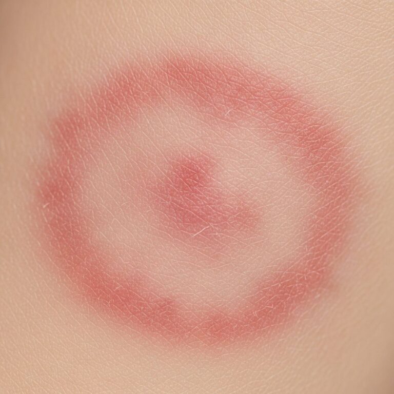
- It typically presents as painful erythematous or purpuric plaques at injection sites that progress to necrosis within days.
- Often associated with heparin-induced thrombocytopenia (HIT), particularly type II HIT, due to IgG antibodies against platelet factor 4 (PF4)–heparin complexes.
- Diagnosis involves clinical suspicion, 4T score, PF4 antibody testing, and skin biopsy showing microvascular thrombosis.
- Management requires immediate heparin discontinuation and switch to non-heparin anticoagulants like direct thrombin inhibitors (e.g., argatroban, bivalirudin) or fondaparinux.
What is heparin-induced skin necrosis?
Heparin-induced skin necrosis (HISN) is an uncommon adverse reaction to heparin anticoagulation therapy. It manifests as areas of skin infarction due to thrombosis in dermal and subcutaneous vessels. While most cases occur at subcutaneous injection sites, distant lesions have been reported, occurring in up to 10-15% of cases.
Heparin, used for prophylaxis and treatment of thromboembolic disorders, binds to antithrombin III to inhibit thrombin and factor Xa. However, in susceptible individuals, it triggers an immune response leading to platelet activation and microthrombi formation. HISN is distinct from other heparin-related skin reactions like delayed-type hypersensitivity (eczema-like plaques) or acute systemic reactions.
Incidence is low: approximately 0.2% with LMWH and 2.6% with unfractionated heparin (UFH), though underreported due to diagnostic challenges. Risk factors include female sex, obesity, diabetes, recent surgery, and prior heparin exposure.
Who gets heparin-induced skin necrosis?
HISN affects patients receiving heparin for various indications:
- Prophylaxis post-surgery (e.g., orthopedic, abdominal).
- Treatment of deep vein thrombosis (DVT), pulmonary embolism (PE), or acute coronary syndrome.
- Therapeutic anticoagulation in pregnancy or atrial fibrillation.
Demographics show predominance in adults aged 40-70 years, with women comprising 60-75% of cases. Those with underlying protein C/S deficiencies or antiphospholipid syndrome may be predisposed, though HIT is the primary driver.
In one series, 50% of patients developed thrombocytopenia, confirming HIT association. Delayed-onset cases up to 30-40 days post-initiation are rare but reported, especially with LMWH like dalteparin or nadroparin.
What causes heparin-induced skin necrosis?
The pathophysiology centers on type II HIT, where polyclonal IgG antibodies bind to PF4-heparin multimolecular complexes on platelet surfaces. This activates FcγRIIa receptors, causing platelet aggregation, microparticle release, and thrombin generation despite thrombocytopenia.
In skin, this leads to endothelial damage, fibrinoid necrosis, and thrombosis of dermal vessels, resulting in ischemia and necrosis. Unlike HIT type I (non-immune, mild platelet drop), type II involves immune-mediated hypercoagulability.
Lesions at distant sites suggest systemic antibody circulation, with microvascular thrombosis independent of local injection trauma. Genetic factors like HLA alleles may increase susceptibility, but evidence is limited.
What are the clinical features of heparin-induced skin necrosis?
Symptoms emerge 5-15 days after heparin initiation (median 7-10 days), though delayed cases occur beyond 30 days. Prodrome includes burning pain or pruritus at sites.
Characteristic progression:
- Day 1-3: Painful, well-demarcated erythematous or violaceous macules/plaques (2-10 cm), often indurated.
- Day 4-7: Central blistering, hemorrhagic bullae, or ecchymosis; surrounding flare.
- Day 7+: Necrosis with black eschar, ulceration, and sloughing of skin/subcutis; full-thickness involvement in severe cases.
Common sites: abdomen (50%), thighs (30%), buttocks. Distant lesions (e.g., limbs, trunk) indicate systemic HIT. Systemic signs: fever (20%), chills; thrombocytopenia in 50-90%; thrombosis (DVT/PE) in 30-50%.

Figure 1: Classic HISN lesion – tender purpuric plaque with central necrosis on thigh.
How is heparin-induced skin necrosis diagnosed?
Diagnosis is clinical, supported by labs and histopathology. High suspicion in heparin-exposed patients with painful necrotic lesions.
4T Score for HIT probability (essential tool):
| Parameter | 0 points | 1 point | 2 points |
|---|---|---|---|
| Thrombocytopenia | <50% fall AND nadir ≥20 | 50-75% fall OR nadir 10-19 | ≥75% fall OR nadir ≤10 OR heparin re-exposure timing |
| Timing | >Day 10 OR ≤1 day (no prior) | Days 5-10 OR ≤1 day (prior) | Day 5-10 (no prior) OR ≤1 day (prior) |
| Thrombosis | None | Progressive/skin necrosis | New thrombosis/necrosis |
| oTher causes | Definite | Possible | None evident |
Score ≥6: high probability; test urgently.
Labs:
- PF4/heparin ELISA (optical density >1.0 high risk).
- Functional assays (SRA, HIPA) for confirmation.
- Platelet count trend; exclude DIC, sepsis.
Histopathology: Dermal hemorrhage, fibrin thrombi in venules/arterioles, endothelial swelling, minimal inflammation – pathognomonic for HIT-related necrosis.
What is the differential diagnosis for heparin-induced skin necrosis?
- Coumadin necrosis: Protein C deficiency; days 3-5, breasts/thighs.
- Infection: Necrotizing fasciitis (rapid spread, systemic toxicity).
- Calciphylaxis: ESRD patients; livedo-like.
- Antiphospholipid syndrome: Livedo, multi-site.
- Hypersensitivity: Eczematous plaques without necrosis.
What is the treatment for heparin-induced skin necrosis?
Immediate actions:
- Discontinue ALL heparin/LMWH – do NOT switch within class.
- Avoid platelet transfusions (worsens thrombosis).
- Initiate non-heparin anticoagulant if thrombosis risk high.
Recommended anticoagulants:
| Agent | Class | Dose example | Notes |
|---|---|---|---|
| Argatroban | Direct thrombin inhibitor | 0.5-2 mcg/kg/min IV | Hepatic clearance; monitor aPTT |
| Bivalirudin | Direct thrombin inhibitor | 0.15-0.2 mg/kg/hr IV | Renal clearance |
| Fondaparinux | Anti-Xa (pentasaccharide) | 2.5 mg SC daily (prophy) | Low HIT cross-reactivity |
| DOACs (apixaban) | Anti-Xa oral | Age/dose adjusted | After platelet recovery |
Wound care: Debridement for full-thickness necrosis; grafts rarely needed. Healing occurs in 2-6 weeks post-discontinuation. Monitor for limb/gangrene-threatening extension.
What is the outcome for heparin-induced skin necrosis?
Prognosis excellent with prompt management: 80-90% full recovery, lesions heal with scarring. Mortality 5-10% from thrombosis/sepsis if delayed. Amegakaryocytic HIT or bilateral adrenal hemorrhage are fatal variants.
Long-term: Lifetime heparin avoidance; document allergy. Fondaparinux safe in most.
Prevention of heparin-induced skin necrosis
- Minimize heparin duration/exposure.
- Monitor platelets q2-3 days from day 4.
- Prefer DOACs over heparin where possible.
- Site rotation; avoid obese areas.
- High-risk patients: baseline 4T, early PF4 testing.
Frequently asked questions about heparin-induced skin necrosis
Q. How soon after starting heparin does skin necrosis appear?
A. Typically 5-15 days; delayed cases up to 30+ days with LMWH.
Q. Is heparin-induced skin necrosis always associated with low platelets?
A. No, only 50% have thrombocytopenia; skin necrosis can be first sign of HIT.
Q. Can I use low-molecular-weight heparin if necrosis occurred with unfractionated?
A. No, cross-reactivity high; switch to non-heparin agents immediately.
Q. What does biopsy show in HISN?
A. Microvascular thrombi, fibrinoid necrosis, minimal inflammation.
Q. Is fondaparinux safe after HISN?
A. Yes, low cross-reactivity; used successfully in cases.
References
- Heparin-Induced Skin Necrosis Occurring at a Distance From … — Actas Dermo-Sifiliográficas. 2020-01-01. https://www.actasdermo.org/en-heparin-induced-skin-necrosis-occurring-at-articulo-S1578219019303026
- Delayed-onset heparin-induced skin necrosis: a rare complication of … — PMC (BMJ Case Reports). 2017-12-01. https://pmc.ncbi.nlm.nih.gov/articles/PMC5747761/
- Skin necrosis and heparin‐induced thrombocytopenia syndrome — PMC (Clinical Case Reports). 2022-01-01. https://pmc.ncbi.nlm.nih.gov/articles/PMC8765090/
- Heparin-Induced Cutaneous Necrosis Unrelated to Injection Sites — JAMA Dermatology. 1990-01-01. https://jamanetwork.com/journals/jamadermatology/fullarticle/544121
- Heparin-induced skin lesions — The Blood Project (PDF). 2022-07-01. https://www.thebloodproject.com/wp-content/uploads/2022/07/HIT_SKIN.pdf
Similar Articles


Read full bio of medha deb