Hibernoma: Symptoms, Diagnosis, And Treatment Guide
Learn about hibernomas: rare benign tumors of brown fat, their diagnosis, and treatment options.

Hibernoma: Overview
A hibernoma is a rare subtype of benign lipomatous tumor derived from brown fat, also known as fetal lipoma or lipoma of embryonic fat. First described in 1906 by German physician H. Merkel under the name ‘pseudolipoma’, hibernomas represent an uncommon neoplasm of brown adipose tissue. Unlike malignant tumors, hibernomas have no association with malignant potential and do not undergo transformation to cancer. These benign soft tissue tumors are characterized by the presence of prominent brown adipocytes that resemble normal brown fat tissue found in hibernating animals.
Clinical Presentation and Symptoms
Hibernomas typically present as slow-growing, painless, solitary masses that develop in the subcutaneous tissues. Most patients remain asymptomatic, with symptoms often present for years before seeking medical attention. The tumors are generally described as:
- Movable and soft in consistency
- Firm to palpation
- Nontender to touch
- Well-defined and mobile masses
- Warm to the touch due to their rich blood supply
While hibernomas are usually painless, they may occasionally cause light pressure or discomfort due to their size. In some cases, the tumor may compress nearby nerves, leading to tenderness or pain. Complications can arise when the growing mass affects surrounding structures, particularly when located in confined anatomical spaces.
Location and Epidemiology
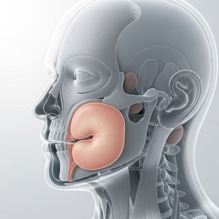
The most common locations for hibernoma include the thigh, shoulder, back, neck, and chest. The inter-scapular region (between the shoulder blades) is the most frequently reported site. Other documented locations include the arm, retroperitoneum, breast, larynx, pleura, pelvis, vulva, and scrotum. Approximately 10% of hibernomas occur in intramuscular tissue, while the vast majority develop in subcutaneous tissues.
Hibernomas generally occur in adults with a peak incidence in the third decade of life, with a predominance in women. They have not been associated with specific age groups beyond this general pattern, and presentation across different age ranges has been documented in clinical literature.
Pathological Characteristics
From a macroscopic perspective, hibernomas appear as well-defined, encapsulated or circumscribed masses with a soft texture. The color typically ranges from yellow-tan to deep brown, depending on lipid concentration. The size of hibernomas varies considerably, typically ranging from 1 to 27 centimeters, with a mean size of approximately 10 centimeters.
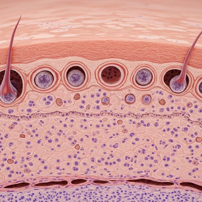
Microscopically, hibernomas are characterized by:
- Large, multi-vacuolated brown fat cells (adipocytes)
- Abundant mature adipose cells within the tissue
- Several small branching capillaries throughout the tumor
- No significant adipocytic atypia (abnormal cell appearance)
- Rich vascularity in the background
- Thin capsules surrounding the lesion
These histological features distinguish hibernomas from standard white fat lipomas and help confirm diagnosis under microscopic examination.
Histological Variants
Four recognized histologic types of hibernomas exist, though one is most frequently observed. The main variants include:
- Typical hibernoma: The most common variant, showing characteristic brown fat cells with the histological features described above
- Lipoma-like hibernoma: Features univacuolated white adipocytes with scattered hibernoma cells, most commonly arising in the thigh region
- Myxoid hibernoma: Contains myxoid stroma along with hibernoma cells and mature adipocytes
- Spindle cell hibernoma: The least common variant, accounting for approximately 2% of hibernomas, featuring spindle cell lipoma characteristics including CD34+ spindle cells, thick collagen bundles, mast cells, and myxoid stroma, typically found on the scalp and posterior neck
Diagnostic Approach
Diagnosis of hibernoma can be challenging because the clinical and radiographic features often resemble both benign lipomas and malignant tumors such as liposarcomas. A thorough medical history and physical examination are essential components of the diagnostic workup.
Key clinical features to assess include:
- The size of the growth
- Whether the mass is movable or fixed
- Any breaks or ulcers on the skin over the mass
- Swellings in regional lymph nodes
- Assessment for additional skin lumps
Important warning signs that may suggest a more serious condition, such as liposarcoma or other malignant tumors, include rapid growth of the mass, pain in specific regions like the armpit or groin suggesting swollen lymph nodes, personal or family history of cancer, and unintended weight loss.
Imaging studies, including non-contrast CT scans, typically show hibernomas as well-demarcated, low-attenuation masses with density relatively close to that of neighboring subcutaneous fat. The size, enhancement pattern, and intramuscular location can sometimes lead to initial misinterpretation as lipoma or liposarcoma. Definitive diagnosis requires histopathological examination, where tissue analysis confirms the presence of brown adipocytes characteristic of hibernoma.
Differential Diagnosis
Several benign and malignant conditions must be differentiated from hibernoma:
Benign conditions that may mimic hibernoma:
- Lipoma and angiolipoma (lipomatous lesions with abundant fatty tissue)
- Fat necrosis (death of fat cells in the body)
- Hemangiomas (collections of blood vessel cells forming a lump)
- Fibromatosis (excessive soft tissue growth)
- Abscess (collection of pus from infection)
Malignant conditions in the differential:
- Liposarcoma (malignant fatty tumor)
- Other soft tissue sarcomas
The clinical indistinguishability between hibernomas and malignant tumors emphasizes the importance of including hibernoma in the differential diagnosis and pursuing definitive histological diagnosis to guide appropriate management.
Prognosis and Complications
Hibernomas are not considered life-threatening tumors. There are no reported cases of malignant transformation, recurrence, or metastatic spread after complete surgical excision. These benign tumors maintain their benign nature throughout the patient’s lifetime when properly managed.
Complications from hibernomas typically arise due to their growing size and effects on surrounding areas. Possible post-operative complications following surgical removal include:
- Delayed wound healing
- Scarring at the surgical site
- Hematoma (collection of blood)
- Seroma (collection of fluid in the tissue)
While hibernomas usually grow slowly and remain asymptomatic, rare presentations have included rapid tumor growth, fever, and weight loss, though these manifestations are uncommon.
Treatment and Management
The primary treatment for symptomatic hibernomas is surgical excision. Complete removal of the tumor is curative and prevents any potential complications from mass effect on adjacent structures. Surgical intervention is particularly recommended when:
- The tumor causes pain or discomfort from nerve compression
- The mass significantly affects quality of life or function
- Patient anxiety is high regarding the lesion
- Rapid growth or concerning symptoms develop
Accurate preoperative diagnosis is critical in reducing patient anxiety and guiding proper surgical strategy. Once completely excised, no surveillance or additional treatment is typically required, as recurrence and malignant transformation have not been documented.
Associated Conditions
While hibernomas are typically sporadic, there has been speculation about an association between hibernomas and multiple endocrine neoplasia type 1 (MEN1). A few case reports have documented hibernoma occurring in patients with MEN1 syndrome, though this association remains uncommon and requires further investigation.
Frequently Asked Questions
What is the difference between a hibernoma and a lipoma?
While both are benign fatty tumors, hibernomas are derived from brown fat and contain characteristic multi-vacuolated brown adipocytes, whereas lipomas are composed of white fat with univacuolated adipocytes. Hibernomas are also rarer and may feel slightly warmer due to their vascular supply.
Can a hibernoma turn into cancer?
No. Hibernomas have no association with malignant potential. There are no reported cases of hibernomas transforming into cancer or spreading to other body parts.
Do all hibernomas require surgery?
Not necessarily. Asymptomatic hibernomas that are not causing functional impairment may be monitored clinically without immediate intervention. Surgery is recommended when the tumor causes symptoms, rapid growth occurs, or patient preference warrants removal.
How is hibernoma definitively diagnosed?
Definitive diagnosis requires histopathological examination of tissue obtained through biopsy or surgical excision, which confirms the presence of characteristic brown adipocytes and brown fat cells.
What is the recurrence rate after surgical removal?
Complete surgical excision of hibernoma is curative with no documented cases of recurrence or metastatic spread after complete removal.
Are there any lifestyle modifications needed after hibernoma diagnosis?
No specific lifestyle modifications are required. Patients with asymptomatic hibernomas can maintain normal activities. Those undergoing surgery should follow standard post-operative care guidelines for wound healing and recovery.
References
- Hibernoma — DermNet NZ. Accessed January 2026. https://dermnetnz.org/topics/hibernoma
- Symptomatic Hibernoma: A Rare Soft Tissue Tumor — National Center for Biotechnology Information (NCBI/PMC). 2012. https://pmc.ncbi.nlm.nih.gov/articles/PMC3525332/
- Hibernoma – StatPearls — NCBI Bookshelf, National Library of Medicine. 2023. https://www.ncbi.nlm.nih.gov/books/NBK570579/
- Hibernoma: What It Is, Causes & Treatment — Cleveland Clinic. Accessed January 2026. https://my.clevelandclinic.org/health/diseases/24149-hibernoma
- Hibernoma — Visual Dx Medical Reference. Accessed January 2026. https://www.visualdx.com/visualdx/diagnosis/hibernoma
- Signs and Symptoms of Hibernoma — MD Searchlight. Accessed January 2026. https://mdsearchlight.com/skin-problems-and-treatments/hibernoma/
Similar Articles


Read full bio of medha deb