Hidradenoma Papilliferum Pathology: An Expert Guide
Comprehensive pathology guide to hidradenoma papilliferum, a benign apocrine gland tumour commonly affecting the vulva.

Hidradenoma papilliferum, also known as papillary hidradenoma, is a rare benign tumour originating from apocrine glands. This lesion typically manifests as a small, solitary dermal or subcutaneous nodule, most frequently located on the vulva in women of reproductive age. Understanding its pathology is crucial for accurate diagnosis and differentiation from malignant counterparts.
Introduction
Hidradenoma papilliferum belongs to the spectrum of sweat gland lesions, specifically those derived from apocrine differentiation. It is predominantly encountered in the anogenital region, particularly the labia majora, but can occasionally appear on the perianal area, nipple, or axilla. Clinically, it presents as a firm, painless, flesh-coloured to red-brown papule or nodule ranging from 0.5 to 2 cm in diameter. The lesion grows slowly and is often asymptomatic unless ulcerated or secondarily infected.
Histologically, it is characterized by a well-defined architecture without epidermal connection, distinguishing it from other adnexal tumours. Its benign nature is confirmed by the absence of atypia, mitoses, or invasion, though rare malignant transformations have been reported. Early recognition prevents unnecessary extensive surgery.
Clinical features
The prototypical presentation is a solitary, well-circumscribed nodule in the vulvar skin. Patients typically report a painless lump noticed incidentally or during self-examination. The overlying skin may appear normal or slightly stretched, with rare surface erosion. In postmenopausal women, hormonal influences may contribute to its development, given frequent positivity for oestrogen and progesterone receptors.
- Location: Vulva (labia majora >90%), perianal, inguinal, rarely extragenital (e.g., eyelid, scalp).
- Size: 0.3–3 cm, average 1 cm.
- Appearance: Dome-shaped papule/nodule, flesh-coloured, pink, or haemorrhagic.
- Symptoms: Asymptomatic; pruritus or discharge if cystic degeneration occurs.
- Associations: Rare links to naevus sebaceus or endocrine disorders.
Biopsy is recommended for any persistent vulvar nodule to exclude malignancy, as clinical diagnosis alone is unreliable.
Pathology
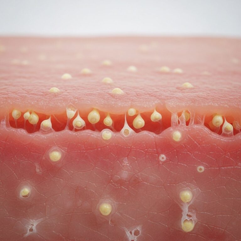
Microscopic examination reveals a well-circumscribed dermal nodule, typically lacking connection to the overlying epidermis (see figure 1). The tumour exhibits an arborizing pattern of tubules and broad, elongated fronds lined by a characteristic double-layered epithelium (see figures 2, 3).
Histology
The inner epithelial layer consists of cuboidal myoepithelial cells, while the outer layer features tall columnar apocrine cells with pale eosinophilic cytoplasm. Decapitation secretion—apical snouting of cytoplasm into the lumen—is a hallmark feature indicative of apocrine differentiation. Larger fronds often contain a central fibrous core, and surrounding fibrous tissue may form a pseudocapsule due to compression.
| Feature | Description |
|---|---|
| Tumour architecture | Well-circumscribed dermal nodule, no epidermal continuity |
| Epithelial lining | Double-layered: inner myoepithelial (cuboidal), outer apocrine (columnar) |
| Cytology | Pale eosinophilic cytoplasm, decapitation secretion |
| Stroma | Fibrous cores in fronds, pseudocapsule |
| Other | No atypia, mitoses rare/absent |
Cystic spaces may form due to dilatation, filled with eosinophilic secretions. PAS staining highlights diastase-resistant granules in apocrine cell apices. No necrosis, lymphovascular invasion, or perineural spread is seen in benign cases.
Immunohistochemistry
Immunoprofile supports apocrine origin:
- Epithelial cells: Positive for keratin (AE1/AE3), EMA, CEA; GCDFP-15 (gross cystic disease fluid protein-15) strongly positive.
- Hormone receptors: Oestrogen receptor (ER), progesterone receptor (PR), androgen receptor (AR) often positive, explaining vulvar predilection.
- Myoepithelial cells: S-100, p63, smooth muscle actin (SMA), calponin positive.
- Other: Negative for CK20, HER2 typically; Ki-67 proliferation index low (<5%).
These markers distinguish it from eccrine tumours (e.g., poroid neoplasms lack GCDFP-15) and carcinomas (high Ki-67, HER2).
Differential diagnosis
Several lesions mimic hidradenoma papilliferum histologically or clinically. Key differentials include:
| Entity | Distinguishing Features |
|---|---|
| Syringocystadenoma papilliferum | Connected to epidermis, crusted/verrucous surface, plasma cell-rich stroma in papillary cores |
| Malignant hidradenoma papilliferum (intraductal ca) | Infiltrative growth, mitoses, atypia, no myoepithelial layer |
| Adenocarcinoma (apocrine/eccrine) | Lacks myoepithelium, marked atypia, invasion, necrosis |
| Hidradenoma (eccrine) | Clear cells, ductal lumina without apocrine secretion, epidermal connection possible |
| Poroma/acrospiroma | Epidermal attachment, poroid/basaloid cells, no fronds |
| Mucinous eccrine carcinoma | Mucin pools, signet-ring cells, infiltrative |
Syringocystadenoma papilliferum, often on scalp associated with naevus sebaceus, shows epidermal invaginations with plasma cell infiltrate—absent in hidradenoma papilliferum.
Management
Complete surgical excision is curative, with low recurrence risk due to its encapsulated nature. Mohs micrographic surgery is ideal for vulvar sites to preserve tissue. No adjuvant therapy needed for benign lesions. Rare malignant variants require wide excision (±sentinel node biopsy) and follow-up.
- Excision margins: 1–3 mm adequate.
- Follow-up: Clinical exam at 6–12 months; monitor for recurrence (rare <5%).
- Malignancy suspicion: If infiltrative/atypical, stage with imaging.
Prognosis
Excellent for benign hidradenoma papilliferum—100% cure post-excision. Malignant transformation is exceptionally rare (<1%), but reported cases show aggressive behaviour akin to hidradenocarcinoma with metastasis potential.
Frequently Asked Questions (FAQs)
Is hidradenoma papilliferum cancerous?
No, it is a benign apocrine tumour. Malignant variants are rare and show invasion/atypia.
What causes hidradenoma papilliferum?
Unknown; hormonal factors implicated due to receptor positivity. No strong genetic links identified.
How is it diagnosed?
Histology post-biopsy/excision; immunohistochemistry confirms apocrine origin.
Does it recur after treatment?
Rarely, if incompletely excised. Recurrence rate <2%.
Can it occur outside the vulva?
Yes, rarely on perianal skin, nipple, or axilla.
References
- Hidradenoma papilliferum pathology — DermNet NZ. 2023. https://dermnetnz.org/topics/hidradenoma-papilliferum-pathology
- Hidradenoma papilliferum pathology image — DermNet NZ. 2023. https://dermnetnz.org/imagedetail/18555-hidradenoma-papilliferum-pathology
- Sweat gland lesions — DermNet NZ. 2023. https://dermnetnz.org/topics/sweat-gland-lesions
- Hidradenocarcinoma pathology — DermNet NZ. 2023. https://dermnetnz.org/topics/hidradenocarcinoma-pathology
- Syringocystadenoma Papilliferum of the Scalp in an Adult — PMC (NCBI). 2013-05-01. https://pmc.ncbi.nlm.nih.gov/articles/PMC3644463/
Similar Articles


Read full bio of medha deb