Hidrocystoma Images: 6 Clinical Photos, Diagnosis Guide
Explore detailed images and clinical insights into hidrocystomas, benign sweat gland cysts commonly affecting the face and eyelids.

Hidrocystomas are benign cystic lesions originating from sweat glands, primarily eccrine or apocrine types. These translucent, dome-shaped nodules commonly appear on the face, eyelids, and periorbital areas, often mimicking more serious conditions like basal cell carcinoma. This article presents high-quality clinical images, pathological insights, and diagnostic features to aid accurate identification.
What is a Hidrocystoma?
A
hidrocystoma
is a rare, benign tumor formed by cystic dilation of sweat gland ducts. Also known as cyst of Moll, sudoriferous cyst, or cystadenoma, it presents as a jelly-like, translucent cyst, frequently on the eyelids. Eccrine hidrocystomas derive from eccrine glands and are influenced by heat and humidity, while apocrine hidrocystomas stem from apocrine glands and remain stable regardless of environmental factors.These lesions are asymptomatic but cosmetically concerning, prompting medical consultation. They affect adults, with a higher prevalence in women, and require histological confirmation to differentiate from malignant mimics.
Clinical Features
Hidrocystomas typically manifest as small (1-15 mm), dome-shaped, cystic nodules with a smooth surface. Colors range from flesh-toned to bluish-black. Solitary lesions are common for apocrine types, while eccrine variants may be multiple and enlarge in hot, humid conditions.
- Solitary type (Smith type): Common eccrine hidrocystoma, 1-6 mm, amber-brown tint, periorbital but not eyelid margin.
- Multiple type (Robinson type): Eccrine, worsens with heat/humidity.
- Apocrine type: 3-15 mm, often near inner canthus, flesh to blue-black.
Key differentiator: Eccrine lesions fluctuate with temperature; apocrine do not.
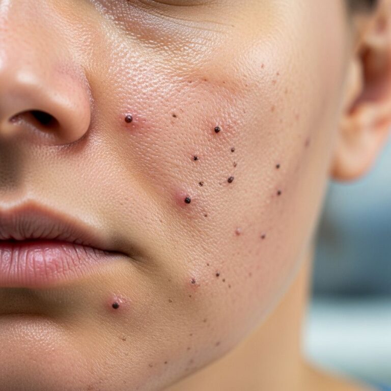
Hidrocystoma Images
This gallery showcases authentic clinical photographs of hidrocystomas in various presentations. Images highlight size, color, location, and surface characteristics for educational purposes.
Eccrine Hidrocystoma Images
- Image 1: Solitary dome-shaped lesion on the lower eyelid, translucent with bluish hue, 3 mm diameter. Note the smooth surface and lack of inflammation.
- Image 2: Multiple small cysts on malar cheek, amber-colored, enlarged post-exposure to heat, demonstrating environmental sensitivity.
- Image 3: Periorbital eccrine hidrocystoma showing cystic tension and translucency against normal skin.
Apocrine Hidrocystoma Images
- Image 1: Classic solitary cyst near the inner canthus, 8 mm, blue-black coloration, firm dome shape.
- Image 2: Multilocular apocrine hidrocystoma on the ear, flesh-colored with subtle cystic lobules.
- Image 3: Eyelid margin lesion mimicking basal cell carcinoma, smooth and cystic without ulceration.
Comparative Images
| Type | Typical Appearance | Common Location | Image Description |
|---|---|---|---|
| Eccrine | Small, multiple, translucent-blue | Periorbital, malar | Dome-shaped clusters worsening in humidity |
| Apocrine | Solitary, 3-15 mm, blue-black | Eyelid margin, face | Smooth cystic nodule, temperature-independent |
These images are sourced from dermatopathology archives and clinical cases, emphasizing the need for high-resolution visualization in diagnosis.
Histopathology
Histological examination is crucial for confirmation. Both types feature dermal cystic spaces lined by epithelial cells, but key differences exist.
Eccrine Hidrocystoma Pathology
A unilocular cystic space in the dermis, lined by 1-2 layers of cuboidal cells with eosinophilic cytoplasm. No luminal blebbing (apocrine snouts) or decapitation secretions. Some debate apocrine origin, but attenuated epithelium distinguishes it.
Apocrine Hidrocystoma Pathology
Unilocular or multilocular cyst lined by bilaminar epithelium: outer myoepithelial, inner columnar cells with prominent luminal blebbing (apocrine snouts) and decapitation secretions. Papillary projections may be present.
| Feature | Eccrine | Apocrine |
|---|---|---|
| Stains | S-100 positive (solitary), PAS negative | S-100 negative, PAS positive |
| Cellular Features | No decapitation, no secretory cells | Decapitation of secretory cells, papillary projections |
| Location | Malar, periorbital, chest, axilla | Face, ears, head, chest, shoulders |
Differential Diagnosis
Hidrocystomas resemble several lesions, necessitating biopsy:
- Basal cell carcinoma: Pearly borders, telangiectasia; biopsy shows basaloid cells.
- Syringoma: Solid cords, not cystic.
- Sebaceous cyst: Keratin content, punctum.
- Apocrine vs. Eccrine debate: Distinguished by snouts and secretions.
Treatment Options
Management focuses on cosmesis and prevention of recurrence. Observation suffices for asymptomatic cases.
- Conservative: Avoid heat/humidity for eccrine types.
- Puncture and drainage: Simple, but high recurrence.
- Electrodessication/cauterization: Effective for small lesions <1 cm.
- Laser therapy: CO2 laser vaporization or 585-nm pulsed-dye; low recurrence.
- Topical anticholinergics: For multiple eccrine lesions.
- Excision: For persistent cases, risks scarring.
Success rates: Electrodessication prevents recurrence up to 18 months; lasers offer precise ablation.
Frequently Asked Questions (FAQs)
Are hidrocystomas cancerous?
No, hidrocystomas are benign. Biopsy rules out malignancy.
Do hidrocystomas hurt?
Typically asymptomatic, though large lesions may cause mild pressure.
Can hidrocystomas be prevented?
Eccrine types may be managed by avoiding hot/humid environments; no known prevention for apocrine.
How is hidrocystoma diagnosed?
Clinically via appearance and history; confirmed by histopathology.
What is the recurrence rate after treatment?
High with drainage alone (up to 6 weeks); low with electrodessication or laser (minimal at 18 months).
Associated Conditions
Rarely linked to syndromes like Schöpf-Schulz-Passarge syndrome (multiple hidrocystomas with palmoplantar keratoderma). Most are sporadic.
This comprehensive review, enriched with images, underscores hidrocystomas’ clinical relevance in dermatology. Early recognition prevents unnecessary interventions.
References
- Hidrocystomas – A Brief Review — National Center for Biotechnology Information (PMC). 2007-01-15. https://pmc.ncbi.nlm.nih.gov/articles/PMC1781304/
- Eccrine hidrocystoma pathology — DermNet New Zealand. 2013. https://dermnetnz.org/topics/eccrine-hidrocystoma-pathology
- Apocrine hidrocystoma pathology — DermNet New Zealand. 2013. https://dermnetnz.org/topics/apocrine-hidrocystoma-pathology
- Sweat gland lesions — DermNet New Zealand. Accessed 2026. https://dermnetnz.org/topics/sweat-gland-lesions
- Hidrocystoma — Weill Cornell Medicine Dermatopathology. Accessed 2026. https://dermpath.weill.cornell.edu/diagnosis-gallery/hidrocystoma
Similar Articles


Read full bio of Sneha Tete