Histiocytosis: Types, Symptoms, Causes & Treatment
Understanding histiocytosis: A comprehensive guide to rare immune system disorders affecting children and adults.

What Is Histiocytosis?
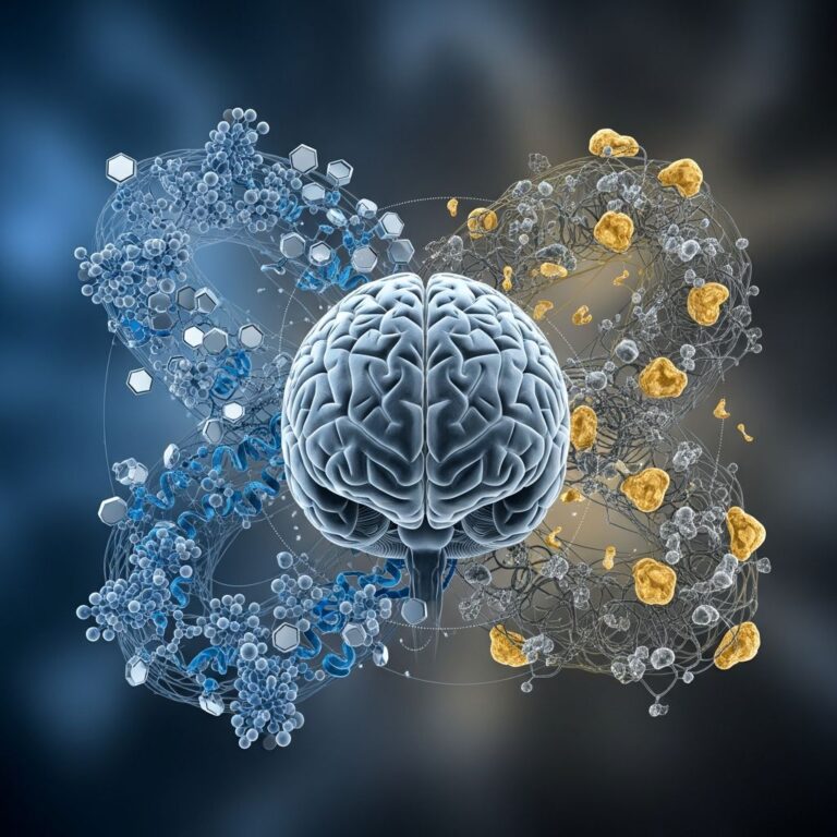
Histiocytosis is a group of rare disorders characterized by an abnormal increase or dysfunction of histiocytes, which are specialized white blood cells that play a crucial role in your immune system. Histiocytes are normally found throughout your body, including in your bone marrow, bloodstream, skin, lungs, spleen, and liver, where they help defend against infections and foreign invaders.
When you have histiocytosis, these immune cells begin to accumulate abnormally in various parts of your body, causing inflammation and tissue damage. The condition can affect people of any age, though certain types are more common in children, while others may develop in adults. Without proper diagnosis and treatment, histiocytosis can lead to serious complications and organ damage.
Histiocytosis belongs to a category of disorders called histiocytic disorders, and it encompasses several distinct subtypes, each with unique characteristics and clinical presentations. The classification and severity of histiocytosis depend on which type you have, where the cells accumulate, and how many body systems are affected.
Types of Histiocytosis
There are several different types of histiocytosis, each with distinct characteristics and clinical features. Understanding the differences between these types is essential for proper diagnosis and treatment planning.
Langerhans Cell Histiocytosis (LCH)
Langerhans cell histiocytosis is a rare disorder that occurs when immature immune system cells called Langerhans cells build up in your child’s or adult’s body. LCH is one of the most common forms of histiocytosis and can affect people of all ages, though it more frequently impacts children and young adults. When Langerhans cells accumulate, they can cause tissue damage and tumor formation in various parts of the body.
LCH is classified as either single-system disease, where one body system is affected, or multi-system disease, where multiple systems are involved. The classification determines the treatment approach and overall prognosis. In certain cases, particularly single-system LCH involving the skin or bone, the disease may improve on its own without treatment, requiring only observation to ensure it doesn’t return or spread.
The severity and outlook depend significantly on which organs are affected. Low-risk organs include the skin, bone, and lymph nodes, while high-risk organs include the liver, spleen, lungs, and bone marrow. When LCH affects high-risk organs, more aggressive treatment approaches are typically necessary.
Rosai-Dorfman Disease (RDD)
Rosai-Dorfman disease, also called sinus histiocytosis with massive lymphadenopathy, is a rare, benign condition involving an overgrowth of histiocytes. RDD is classified as a form of non-Langerhans cell histiocytosis and typically presents with enlarged lymph nodes in the neck (lymphadenopathy), though excess histiocytes can affect other lymph nodes and body regions as well.
RDD mainly affects children, teens, and young adults, with most diagnoses occurring around age 20. However, documented cases of RDD have been recorded in people in their 70s. The condition looks and behaves differently in different individuals, with cells potentially affecting one group of lymph nodes or multiple groups. About 40% of people with RDD have excess histiocytes in extranodal sites—areas outside the lymph nodes—in addition to their enlarged lymph nodes.
The benign nature of RDD is a key distinction from other histiocytosis types, and many patients experience mild symptoms or may have no symptoms at all if the histiocytes only affect lymph nodes. However, if excess histiocytes prevent an organ from functioning correctly, more severe symptoms can develop.
Erdheim-Chester Disease (ECD)
Erdheim-Chester disease is a rare blood disorder that occurs when your body makes too many white blood cells called histiocytes. ECD can affect multiple organs and cause various symptoms or even no symptoms, making diagnosis particularly challenging. Unlike RDD, ECD can be more aggressive and may cause significant tissue and organ damage if left untreated.
With ECD, histiocytes grow out of control and may travel to different parts of your body where they’re not usually found, causing tumors and tissue invasion. Scientists have discovered that over half of people with ECD have a mutation in the BRAF gene that promotes uncontrolled histiocyte growth. Although BRAF is the most common site for gene mutations with ECD, other gene mutations have also been discovered, and these genetic discoveries have allowed scientists to develop targeted treatments that prevent abnormal histiocyte growth.
Hemophagocytic Lymphohistiocytosis (HLH)
Hemophagocytic lymphohistiocytosis is a rare and often life-threatening condition if left untreated. HLH causes your immune system to attack your body instead of a foreign invader like a virus. An infection can trigger your immune system to produce too many cells, which instead of fighting the infection, attack your body and make you even more ill.
HLH is either acquired or genetic, and there’s no way to prevent it. The condition occurs when your immune system abnormally overreacts, leading to an overproduction of immune system cells called histiocytes and T cells, which attack your body instead of attacking foreign invaders. These white blood cells don’t have the correct instructions in your DNA to perform their jobs as they should, causing the symptoms of HLH.
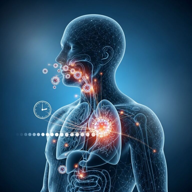
Symptoms of Histiocytosis
The symptoms of histiocytosis vary significantly depending on the type you have, where the excess histiocytes accumulate in your body, and which organs or systems are affected. Some individuals may experience mild symptoms or even no symptoms, while others may have severe manifestations requiring immediate medical attention.
Common Symptoms
For Rosai-Dorfman disease, the most common symptom is painless, swollen lumps on both sides of your neck, as excess histiocytes typically grow in the lymph nodes in this area. Swelling may appear in another location depending on which lymph nodes are affected. If you have only lymph node involvement, you may experience mild symptoms or no symptoms at all.
With Erdheim-Chester disease, symptoms depend on which organs are damaged and how severely. Up to half of people with ECD are diagnosed with diabetes insipidus, which causes symptoms like frequent urination and increased thirst due to damage to the pituitary gland. The most common sign of ECD on the skin is yellowish growths on the eyelids, though yellowish-brown growths may also appear on other areas of the body.
Organ-Specific Symptoms
When histiocytes damage glands that release hormones regulating important body processes, various hormone-related symptoms can develop. Excess histiocytes can damage tissue in the brain and nervous system, potentially causing neurological symptoms. ECD affecting one or both eyes can cause vision-related symptoms, while excess histiocytes affecting the lungs often appear on imaging and may cause pulmonary symptoms if severe.
Left untreated, histiocytosis affecting the lungs can cause serious, long-term lung scarring known as pulmonary fibrosis. When histiocytes affect the heart and blood vessels, the damage may be life-threatening without treatment, potentially causing cardiovascular complications. Excess histiocytes can also collect in the spleen, liver, and bone marrow, causing tissue damage and organ dysfunction.
Life-Threatening Signs
In hemophagocytic lymphohistiocytosis, life-threatening signs require immediate emergency care. If you experience any of these critical symptoms, you should visit the emergency room immediately, as early intervention can be lifesaving.
Causes of Histiocytosis
The causes of histiocytosis vary depending on the specific type of the disorder. An overactive or irregularly functioning immune system causes hemophagocytic lymphohistiocytosis, with the condition being either acquired or genetic. Scientists aren’t completely certain what causes the out-of-control cell growth in all instances of other histiocytosis types, but they’ve recently discovered gene mutations that likely play a significant role.
For Erdheim-Chester disease specifically, more than half of people have a mutation in the BRAF gene that promotes uncontrolled histiocyte growth. Although BRAF is the most common site for gene mutations with ECD, scientists have also discovered other gene mutations associated with the disease. These genetic discoveries have revolutionized understanding of histiocytosis and allowed scientists to develop treatments that target specific mutations and prevent abnormal histiocyte growth.
With Rosai-Dorfman disease, scientists don’t know what causes the condition. RDD affects people so differently that it likely has multiple causes. Recent research suggests that cutaneous RDD (CRDD) likely has different causes than classic RDD, though the specific mechanisms remain under investigation.
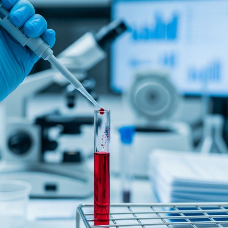
Diagnosis of Histiocytosis
Diagnosing histiocytosis can be challenging because symptoms vary widely from person to person and may mimic other conditions. Healthcare providers typically use a combination of physical examination, imaging studies, and tissue samples to confirm histiocytosis diagnosis.
The diagnostic process usually begins with a thorough medical history and physical examination. Imaging tests such as X-rays, CT scans, or MRI may be performed to identify affected areas and assess the extent of disease involvement. A definitive diagnosis typically requires a tissue biopsy, where a small sample of affected tissue is examined under a microscope to identify the characteristic histiocyte accumulation. For some types, genetic testing may be recommended to identify specific mutations that can guide targeted treatment decisions.
Treatment Options
Treatment approaches for histiocytosis depend on several factors, including the specific type of histiocytosis, how many and which organs are affected, disease severity, and how the disease is progressing.
Observation and Monitoring
In certain cases, particularly single-system LCH involving the skin or bone, the disease may improve on its own without treatment. In these instances, treatment involves observation to ensure the disease doesn’t return or spread. Regular follow-up appointments and imaging studies help monitor disease status without exposing patients to unnecessary medications or procedures.
Targeted Therapy and Immunotherapy
Current treatments for ECD include targeted therapy, immunotherapy, and chemotherapy. Targeted therapies work by specifically blocking the abnormal genetic mutations, particularly BRAF mutations, that drive histiocyte growth. These treatments have significantly improved outcomes and often have fewer side effects compared to traditional chemotherapy.
Combination Approaches
Many patients require combination treatment approaches tailored to their specific disease characteristics. Healthcare providers may combine different treatment modalities based on disease extent, affected organs, and individual patient factors to optimize outcomes and minimize side effects.
Prognosis and Outlook
The prognosis for histiocytosis varies considerably depending on multiple factors. For Rosai-Dorfman disease, prognosis depends on various factors, including how many lymph nodes are affected, where the excess histiocytes are in your body, and your response to treatment. Often, RDD resolves on its own, though in other instances it may require treatment or could cause serious complications.
For Langerhans cell histiocytosis, the outlook depends significantly on where the cells affect the body and whether single or multiple organ systems are involved. Children with single-system disease affecting low-risk organs generally have excellent prognoses, while those with multi-system disease affecting high-risk organs may require more aggressive treatment and have more guarded outcomes.
With early diagnosis and appropriate treatment, most people with histiocytosis can achieve good outcomes and long-term survival. However, left untreated, some forms of histiocytosis can be life-threatening and cause serious complications.
Frequently Asked Questions
Q: Is histiocytosis cancer?
A: Langerhans cell histiocytosis is considered an inflammatory neoplasm—technically neither an autoimmune condition nor cancer. However, the condition requires treatment to prevent tissue damage and complications.
Q: Can histiocytosis be prevented?
A: There is no known way to prevent histiocytosis. Both genetic and acquired forms exist, and prevention strategies are not currently available. However, early diagnosis and prompt treatment can significantly improve outcomes.
Q: How is histiocytosis diagnosed?
A: Diagnosis typically involves a combination of physical examination, imaging studies (CT scans, MRI, X-rays), and tissue biopsy. For some types, genetic testing may be performed to identify specific mutations.
Q: What is the difference between Langerhans cell and non-Langerhans cell histiocytosis?
A: Langerhans cell histiocytosis involves abnormal accumulation of Langerhans cells specifically, while non-Langerhans cell histiocytosis involves other types of histiocytes, such as in Rosai-Dorfman disease and Erdheim-Chester disease.
Q: Can histiocytosis resolve without treatment?
A: In certain cases, particularly single-system Langerhans cell histiocytosis affecting the skin or bone, the disease may resolve on its own. However, most cases require monitoring and many require active treatment to prevent complications.
Q: What are the long-term complications of untreated histiocytosis?
A: Untreated histiocytosis can cause serious complications including organ damage, pulmonary fibrosis in the lungs, cardiac complications, neurological damage, and in some cases, life-threatening conditions requiring immediate emergency care.
References
- Hemophagocytic Lymphohistiocytosis: Symptoms, Causes & Outlook — Cleveland Clinic. https://my.clevelandclinic.org/health/diseases/24292-hemophagocytic-lymphohistiocytosis
- Erdheim-Chester Disease (ECD): Symptoms and Treatment — Cleveland Clinic. https://my.clevelandclinic.org/health/diseases/24668-erdheim-chester-disease
- Rosai-Dorfman Disease: Symptoms, Types & Treatment — Cleveland Clinic. https://my.clevelandclinic.org/health/diseases/24145-rosai-dorfman-disease
- Langerhans Cell Histiocytosis – Cleveland Clinic — Cleveland Clinic. https://my.clevelandclinic.org/health/diseases/17156-langerhans-cell-histiocytosis
- Generalized Non-Langerhans Cell Histiocytosis: Four Cases Illustrating Overlap Among Subtypes — Pennsylvania State University. https://pure.psu.edu/en/publications/generalized-nonlangerhans-cell-histiocytosis-four-cases-illustrat/
- Get Langerhans Cell Histiocytosis Treatment — Cleveland Clinic. https://my.clevelandclinic.org/services/langerhans-cell-histiocytosis-treatment
- Incidence, Clinical Features, and Outcomes of Langerhans Cell Histiocytosis — PubMed/National Center for Biotechnology Information. https://pubmed.ncbi.nlm.nih.gov/35082244/
Similar Articles


Read full bio of medha deb