Histiocytosis: What To Know About Symptoms, Diagnosis, Treatment
Comprehensive guide to histiocytosis: types, symptoms, diagnosis, and treatments for this rare group of disorders.

Histiocytosis refers to a diverse group of rare disorders characterized by the abnormal proliferation and accumulation of
histiocytes
, which are specialized white blood cells derived from the monocyte-macrophage lineage. These cells normally function in immune surveillance and tissue repair but, in histiocytosis, infiltrate various organs, leading to inflammation, tissue damage, and clinical manifestations ranging from benign skin lesions to life-threatening multisystem disease. The term encompasses both neoplastic (clonal proliferation driven by genetic mutations) and reactive (non-clonal, inflammatory) processes, primarily classified intoLangerhans cell histiocytosis (LCH)
andnon-Langerhans histiocytoses (NLCH)
based on cellular characteristics, immunophenotype, and molecular features.Clinically, histiocytosis most commonly presents in children but can affect adults across all ages. Skin involvement occurs in up to 50% of cases, often as the initial manifestation, with lesions mimicking infections, eczema, or malignancies. Bone lesions, particularly in LCH, appear as lytic ‘punched-out’ defects on imaging. Multisystem disease may involve the pituitary (causing diabetes insipidus), lungs, liver, spleen, and central nervous system, necessitating multidisciplinary management. Recent advances in genomic profiling have revealed MAPK/ERK pathway mutations (e.g., BRAF V600E in 50-60% of LCH cases), transforming histiocytosis from an enigmatic syndrome to a targeted therapy-responsive neoplasm. Prognosis varies widely: single-system disease often regresses spontaneously, while risk-organ involvement (liver, spleen, bone marrow) predicts poorer outcomes without prompt intervention.
What is histiocytosis?
Histiocytes are tissue-resident macrophages and dendritic cells pivotal in antigen presentation and phagocytosis. In histiocytosis, their dysregulated accumulation disrupts normal tissue architecture. The 2022 Histiocyte Society classification delineates five groups: LCH, Erdheim-Chester disease (ECD), Rosai-Dorfman disease (RDD), juvenile xanthogranuloma (JXG) family, and indeterminate dendritic cell tumor, reflecting distinct ontogeny and driver mutations. LCH features CD1a+ and Langerin+ (CD207+) cells with Birbeck granules on electron microscopy, distinguishing it from NLCH, which lacks these markers but expresses CD68 and CD163. Incidence is approximately 5-9 per million children yearly for LCH, rarer in adults. Pathophysiology involves somatic mutations triggering cytokine storms and osteoclast activation, explaining osteolytic lesions and chronic inflammation.
Who gets histiocytosis?
Histiocytosis predominantly affects children under 15 years, with LCH peaking at ages 1-3 years. ECD and RDD favor adults (median age 50-60). Pediatric cases often present as multisystem LCH with risk-organ involvement in 10-20%, while adult disease is typically single-system or NLCH. Genetic predisposition is minimal; however, familial cases occur in <5%. No strong ethnic bias exists, though reporting disparities may influence epidemiology. High-risk features include infants under 1 year and multisystem disease.
What causes histiocytosis?
Most cases arise sporadically due to somatic mutations in myeloid precursors. BRAF V600E mutation drives 50-60% of LCH and 20-40% of ECD, activating MAPK signaling and promoting survival/proliferation. Other mutations include MAP2K1 (15-20%), ALK fusions (rare), and NRAS. Reactive histiocytosis (e.g., sinus histiocytosis) links to infections or autoimmunity without clonality. Environmental triggers like smoking exacerbate pulmonary NLCH. These insights enable precision medicine, with BRAF inhibitors (vemurafenib) yielding >80% response rates in mutated cases.
What are the clinical features of histiocytosis?
Manifestations depend on histiocyte distribution:
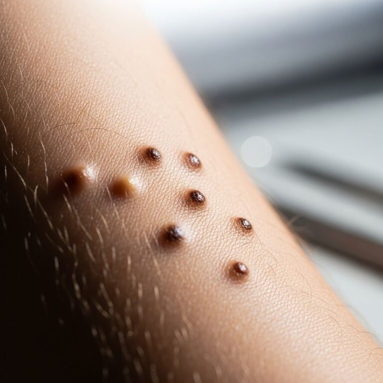
- Skin: Seborrhoeic-like dermatitis, papules, vesicles, ulcers, or nodules; scalp ‘cradle cap’ common in infantile LCH.
- Bone: Painful lytic lesions (skull, long bones, vertebrae); pathological fractures; mastoiditis causing chronic otorrhea.
- Pituitary: Central diabetes insipidus (30% LCH cases); thirst/polyuria.
- Neurologic: Headaches, ataxia, exophthalmos, cognitive deficits from cerebellar/brainstem lesions.
- Pulmonary: Dyspnea, pneumothorax in adult smokers.
- Gastrointestinal/Hepatic: Jaundice, ascites, diarrhea from sclerosing cholangitis.
- Hematologic: Anemia, thrombocytopenia from bone marrow infiltration.
Systemic symptoms include fever, weight loss, irritability.
How is histiocytosis diagnosed?
Diagnosis mandates biopsy with immunohistochemistry (IHC). For LCH: CD1a+, S100+, langerin+; Birbeck granules confirmatory. NLCH shows CD68+/CD163+ without CD1a. Imaging includes skeletal survey, MRI (head/orbits), chest CT, PET-CT for staging. Laboratory: CBC, liver enzymes, ESR/CRP, pituitary function (ADH, TSH). Bone marrow biopsy if pancytopenia. Mutation testing (BRAF, NGS panels) guides therapy. Differential includes lymphoma, infections (TB), sarcoidosis, juvenile xanthogranuloma.
| Symptom | Causes | Recommended Tests |
|---|---|---|
| Thirst, polyuria | Diabetes insipidus | Head MRI, urine/plasma osmolality |
| Bone pain, limping | Osteolytic lesions | X-ray, MRI, skeletal survey |
| Rash/scalp scaling | Skin infiltration | Skin biopsy |
| Headache, seizures | CNS involvement | Brain MRI, neurology exam |
| Jaundice, hepatomegaly | Liver infiltration | LFTs, USG, biopsy |
What is the treatment of histiocytosis?
Treatment stratifies by extent: observation for regressing single-system; chemotherapy for multisystem. Vinblastine + prednisone (standard LCH induction); maintenance with 6-mercaptopurine, methotrexate. Targeted: BRAF inhibitors for V600E+ (cobimetinib, vemurafenib). Surgery/radiation for accessible lesions (e.g., bone curettage). Immunosuppressants (cladribine, cytarabine) for refractory disease. Supportive: desmopressin for DI, hormone replacement. Clinical trials explore JAK inhibitors, anti-CSF1R. Prognosis: >90% cure for low-risk LCH; 60-70% for high-risk.
Complications of histiocytosis
Acute: organ failure (liver, lung). Chronic: endocrinopathies (growth failure, hypogonadism), neurodegeneration (ataxia, dementia in ECD), secondary malignancies, sclerosing cholangitis. Pulmonary fibrosis in adults; fractures from osteolysis.
Prevention of histiocytosis
No known prevention; genetic counseling for rare familial clusters. Smoking cessation reduces NLCH risk.
Timeline of histiocytosis
- Week 1-4: Diagnosis via biopsy/IHC/imaging.
- Month 1-6: Induction chemotherapy; response assessment.
- Year 1-2: Maintenance; monitor reactivation (20% risk).
- Long-term: Endocrine surveillance, annual MRI.
Patient outcomes for histiocytosis
Single-system: 95% resolution. Multisystem low-risk: 80-90% cure. High-risk: 50-70% survival with intensive therapy. Relapse in 30%; neurodegeneration in 10-20% CNS+ cases.
Frequently asked questions about histiocytosis
Q: Is histiocytosis cancer?
A: LCH behaves as a myeloid neoplasm due to clonality/mutations but is often curable unlike solid cancers.
Q: Can histiocytosis be cured?
A: Yes, in >80% low-risk cases; high-risk requires prolonged therapy.
Q: What is the prognosis for children with LCH?
A: Excellent for single-system (95%); poorer if risk organs involved.
Q: Does histiocytosis spread?
A: Not metastatic like cancer; multifocal due to circulating precursors.
Q: Can adults get Langerhans cell histiocytosis?
A: Yes, typically single-system pulmonary/skin.
References
- Histiocytosis Treatment — Memorial Sloan Kettering Cancer Center. 2023. https://www.mskcc.org/cancer-care/types/histiocytosis/histiocytosis-treatment
- Diagnosis and treatment of Langerhans Cell Histiocytosis with bone lesion — PMC / NIH. 2019-08-28. https://pmc.ncbi.nlm.nih.gov/articles/PMC6702438/
- Langerhans cell histiocytosis Treatment — St. Jude Children’s Research Hospital. 2024. https://www.stjude.org/care-treatment/treatment/immune-disorders/histiocytosis/langerhans-cell-histiocytosis.html
- Langerhans Cell Histiocytosis Diagnosis & Treatment — Columbia Neurosurgery. 2023. https://www.neurosurgery.columbia.edu/patient-care/conditions/langerhans-cell-histiocytosis
- Histiocytosis, Immune Disorder — Tampa General Hospital. 2024. https://www.tgh.org/institutes-and-services/conditions/histiocytosis
- Langerhans cell histiocytosis — Cincinnati Children’s Hospital. 2024. https://www.cincinnatichildrens.org/health/l/langerhans-cell-histiocytosis-lch
- Langerhans Cell Histiocytosis Treatment (PDQ®)–Patient Version — National Cancer Institute. 2025-01-10. https://www.cancer.gov/types/langerhans/patient/langerhans-treatment-pdq
Similar Articles


Read full bio of medha deb