Histological Clearance and Recurrence of Keratinocyte Cancers
Understanding histological clearance, recurrence risks, and follow-up strategies for keratinocyte cancers like BCC and SCC.

Keratinocyte cancers, comprising basal cell carcinoma (BCC) and cutaneous squamous cell carcinoma (cSCC), represent the most common malignancies in fair-skinned populations, primarily driven by ultraviolet radiation (UVR) exposure. Histological clearance—confirmed complete removal of tumor cells at excision margins—is the gold standard for assessing treatment success and predicting recurrence risk. This article examines the pathology, risk factors, treatment modalities ensuring clearance, recurrence predictors, and tailored follow-up protocols.
What are Keratinocyte Cancers?
Keratinocyte carcinoma originates in the epidermis, the skin’s outermost layer, from keratinocytes—the predominant epidermal cells. The two primary subtypes are:
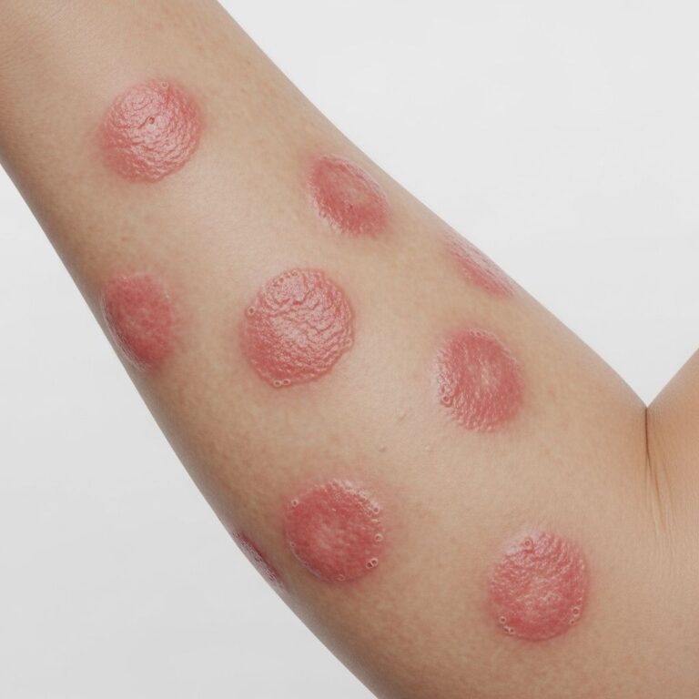
- Basal cell carcinoma (BCC): Arises from basal keratinocytes in the lower epidermis. Typically presents as shiny, pearly nodules with telangiectasia, often on sun-exposed areas like the head and neck.
- Cutaneous squamous cell carcinoma (cSCC): Develops from epidermal keratinocytes or hair follicles, appearing as indurated, nodular, keratinizing, or crusted tumors that may ulcerate. Lifetime risk is 5-15%.
UVR is the primary carcinogen, inducing C>T or CC>TT mutations (Signature 7) with high mutational burden. Additional risks include immunosuppression, aging, and pigmentary factors.
Histological Clearance: Definition and Importance
Histological clearance means no residual tumor cells at surgical margins on microscopic examination post-excision. It is essential for:
- Confirming complete tumor removal.
- Stratifying recurrence and metastasis risk.
- Guiding follow-up frequency.
Without clearance, recurrence rates rise significantly. For low-risk tumors with clearance, recurrence is <5%; high-risk uncleared cases exceed 20%. Biopsies must include deep reticular dermis to detect subclinical invasion.
Pathophysiology and Risk Factors for Recurrence
Recurrence stems from incomplete excision, aggressive subtypes, or host factors. Key histological recurrence predictors include:
| Risk Factor | BCC | cSCC |
|---|---|---|
| Tumor thickness/depth | >2mm or Clark IV/V | >2mm or IV/V |
| Differentiation | N/A | Poorly differentiated |
| Subtypes | Morpheaform, infiltrative | Adenoid, desmoplastic, metaplastic |
| Perineural invasion | Yes | Yes |
| Margins | Involved | Involved |
Patient factors: Immunosuppression (e.g., transplant recipients) increases risk via impaired immune surveillance; HLA mismatch may protect. UVR history, prior skin cancers, and site (H-zone face highest risk) amplify recurrence.
Treatment Options Ensuring Histological Clearance
Treatment prioritizes complete excision with margin confirmation.
Surgical Excision
Gold standard: Complete excision with histological margin assessment.
- Low-risk BCC (<2cm, superficial): 4mm margin.
- High-risk BCC: Mohs micrographic surgery for 99% clearance.
- cSCC: 6mm margin; wider for high-risk.
Non-Surgical Options (Histology-Limited)
- Curettage + electrodessication: Superficial BCC/cSCC <2cm.
- Imiquimod 5% cream: Nodular BCC, Bowen’s disease.
- Cryotherapy: Confirmed superficial lesions with follow-up.
- Radiotherapy: Non-surgical candidates.
Post-treatment histology dictates clearance status.
Follow-Up After Treatment
Frequency hinges on clearance and risk.
| Risk Level | Clearance Status | Follow-Up |
|---|---|---|
| Low | Clear | 6-12 months, then annually |
| High | Clear | 3-6 months x2, then 6-12 months |
| Any | Incomplete | 3 months, re-excision |
| Multiple prior KCs | N/A | Lifelong 6-monthly |
Monitor for recurrence, new primaries, metastasis. High-risk: Imaging if deep invasion.
High-Risk Features and Aggressive Subtypes
High-risk elevates recurrence/metastasis:
- Location: H-zone (central face, ears, nose).
- Size: >2cm.
- Invasion: Bone, nerve, vessel.
- Immunosuppression: 65-250x BCC risk, 100x cSCC.
Aggressive cSCC: Adenosquamous, desmoplastic. βHPV may indirectly contribute via DNA repair inhibition.
Prevention and Emerging Strategies
Sun protection reduces incidence. For high-risk, topical therapies and immunotherapies target residual disease. Genomic analyses identify susceptibility genes for early intervention.
Frequently Asked Questions (FAQs)
What does histological clearance mean for skin cancer?
Complete absence of tumor cells at excision margins on pathology, confirming successful treatment.
How often should patients with keratinocyte cancer be followed up?
Depends on clearance and risk: 3-6 months for high-risk/clear, more frequent if incomplete; lifelong for multiples.
What are the main risk factors for recurrence?
Incomplete margins, high-risk subtypes, immunosuppression, UV exposure history.
Is Mohs surgery always needed for BCC?
No, reserved for high-risk (facial, recurrent, morpheaform); standard excision suffices for low-risk.
Can keratinocyte cancers metastasize?
BCC rarely (<0.1%); cSCC 2-5%, higher in high-risk (poor differentiation, perineural).
Conclusion
Achieving histological clearance minimizes recurrence in keratinocyte cancers. Risk-stratified treatment and vigilant follow-up optimize outcomes, with UV protection preventing new lesions.
References
- Melanoma and keratinocyte cancers – SunSmart GP Guide — Cancer Council Victoria. 2023. https://www.sunsmart.com.au/downloads/resources/booklets/melanoma-other-skin-cancers-gp-guide.pdf
- Squamous Cell Carcinoma (SCC): Guidelines, Diagnosis, & Treatment — The Plastics Fella. 2024. https://www.theplasticsfella.com/squamous-cell-carcinoma/
- Keratinocyte Carcinomas: Current concepts and future research — National Center for Biotechnology Information (PMC). 2019-04-01. https://pmc.ncbi.nlm.nih.gov/articles/PMC6467785/
- General practice management of keratinocyte cancer — Cancer Council Australia. 2023. https://www.cancer.org.au/assets/pdf/quick-reference-guide-general-practice-management-of-keratinocyte-cancer
- Basal Cell and Cutaneous Squamous Cell Carcinomas — American Academy of Family Physicians (AAFP). 2020-09-15. https://www.aafp.org/pubs/afp/issues/2020/0915/p339.html
- Pathogenesis of Keratinocyte Carcinomas and the Therapeutic Implications — National Center for Biotechnology Information (PMC). 2021. https://pmc.ncbi.nlm.nih.gov/articles/PMC8037492/
- Definition of keratinocyte carcinoma — National Cancer Institute (NCI). 2024. https://www.cancer.gov/publications/dictionaries/cancer-terms/def/keratinocyte-carcinoma
Similar Articles


Read full bio of medha deb