Homonymous Hemianopsia: Causes, Symptoms & Treatment
Understanding homonymous hemianopsia: visual field loss, diagnosis, and rehabilitation strategies.

What is Homonymous Hemianopsia?
Homonymous hemianopsia (HH), also referred to as hemianopia, is a visual field deficit that affects the same side of the visual field in both eyes. This means a person loses vision in the right half or left half of their visual world in both eyes simultaneously. Unlike other vision problems that affect only one eye, homonymous hemianopsia impacts corresponding halves of both eyes due to damage along the visual pathway in the brain. This condition often results from cerebrovascular injury, traumatic brain injury, or other neurological events affecting the posterior visual pathways.
The term “homonymous” refers to the fact that both eyes are affected in the same location of their visual fields, while “hemianopsia” literally means “half vision loss.” This distinction is important because it helps healthcare providers identify where along the visual pathway the damage has occurred.
Causes of Homonymous Hemianopsia
Homonymous hemianopsia results from damage or injury to specific brain regions responsible for processing visual information. The visual pathway extends from the eyes through the optic nerves, optic chiasm, optic tract, lateral geniculate nucleus, optic radiations, and finally to the visual cortex in the occipital lobe. Damage at any point along this pathway can result in hemianopsia.
Primary Causes
Stroke remains the most common cause of homonymous hemianopsia. When a stroke occurs, it can damage blood circulation to brain regions responsible for vision processing, including the occipital lobe, optic radiations, or optic tract. A significant proportion of patients develop this condition following cerebrovascular accidents.
Traumatic Brain Injury (TBI) is another major cause. Any damage to the head or brain can impair the visual pathways, resulting in hemianopsia symptoms. This may occur from motor vehicle accidents, falls, assaults, or sports-related injuries.
Brain Tumors and growths can cause homonymous hemianopsia by pushing on or damaging the optical cortex or visual pathways. The location and size of the tumor determine the extent of visual field loss.
Multiple Sclerosis (MS) lesions can disrupt the neural pathways associated with vision. Since MS affects the myelin sheath protecting nerve fibers, it can damage the optic radiations and other visual pathway components.
Infections and Inflammations such as encephalitis or meningitis can contribute to homonymous hemianopsia by affecting brain tissue and disrupting visual processing regions.
Brain Surgery conducted around visual regions may sometimes induce hemianopsia as a complication, creating visual field deficiency symptoms.
Transient or Temporary Causes
Some cases of homonymous hemianopsia are temporary and resolve spontaneously. These include:
– Transient ischemic attacks (TIAs)
– Migraine with visual symptoms
– Occipital, temporal, or parietal lobe seizures
– Nonketotic hyperglycemia
Location of Brain Lesions
Research shows that lesions causing homonymous hemianopsia occur at varying locations:
– Occipital lobe: 45%
– Optic radiation: 32.2%
– Combination of multiple areas: 11.4%
– Optic tract: 10.2%
– Lateral geniculate nucleus: 1.3%
Symptoms and Signs
Recognizing the symptoms of homonymous hemianopsia is crucial for early diagnosis and treatment. Patients typically experience various manifestations affecting their daily functioning and quality of life.
Common Symptoms
Partial or complete loss of vision in the same side of both eyes is the hallmark symptom. Patients may describe this as sudden blindness affecting half their visual field.
Difficulty reading and navigating spaces occurs because patients cannot see text or obstacles on the affected side. Reading becomes challenging as they cannot track lines of text or see the beginning or end of sentences.
Frequent bumping into objects due to reduced peripheral vision on the affected side is common. Patients may collide with furniture, doorways, or people positioned on their blind side.
Unexplained falls or disorientation in familiar surroundings may occur, especially when navigating environments where the affected side is relevant. Patients lose awareness of their environment on the blind side.
Difficulty with road crossing and driving safely becomes problematic, as patients cannot monitor traffic approaching from the blind side.
Increased head and eye movements occur as patients attempt to compensate for their visual field loss by turning their heads more frequently to scan their environment.
Associated Neurological Symptoms
In approximately 53.5% of patients, homonymous hemianopsia appears alongside other neurological symptoms. These may include motor symptoms such as weakness or numbness of the face, arm, or leg; cognitive changes; or a combination of both. Some patients may also experience dizziness, confusion, speech difficulties, or loss of consciousness, particularly if the underlying cause is a stroke.
Diagnosis of Homonymous Hemianopsia
Accurate diagnosis requires a comprehensive evaluation combining clinical examination with specialized testing and neuroimaging.
Clinical Examination
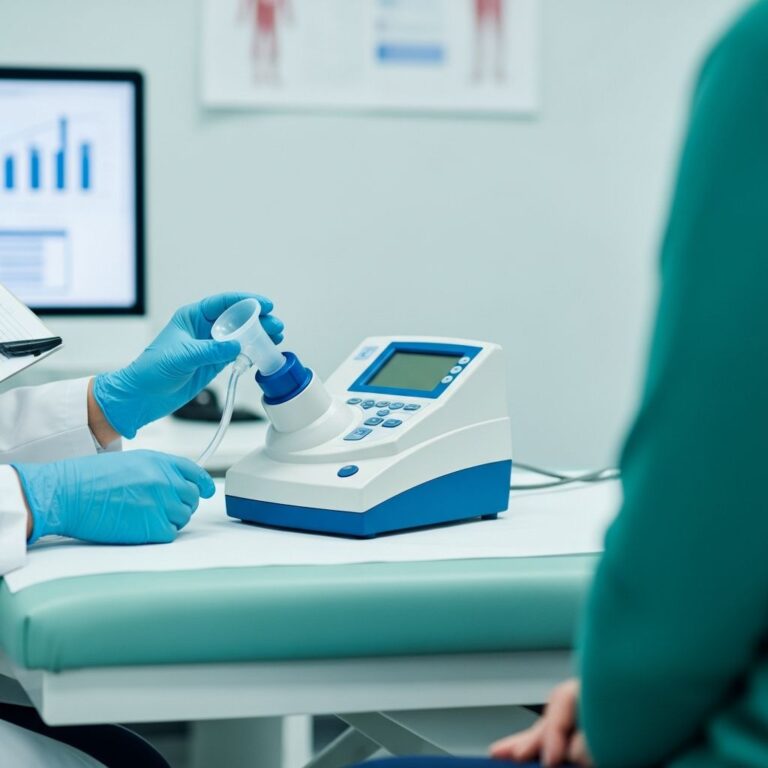
All patients presenting with suspected homonymous hemianopsia should receive a comprehensive medical eye evaluation. This includes assessment of visual acuity, pupillary reaction, intraocular pressure measurement, slit-lamp examination, and fundus examination. These basic assessments help rule out other eye conditions and establish baseline visual function.
Confrontation perimetry is a useful bedside method to detect homonymous hemianopsia. During this test, the examiner sits facing the patient and moves their fingers in different areas of the visual field while the patient maintains a fixed gaze. The patient indicates when they see the movement, helping identify visual field defects.
Neurological examination for localizing signs is crucial for determining the location of the lesion. Specific patterns of visual field loss can help localize the damage site along the visual pathway.
Neuroimaging Studies
All patients experiencing homonymous hemianopsia should undergo advanced neuroimaging to identify the underlying cause. Magnetic resonance imaging (MRI) or computed tomography (CT) scans of the brain reveal structural abnormalities, lesions, infarcts, or tumors responsible for the visual field loss.
MRI typically provides superior detail for visualizing brain tissue and is often preferred for evaluating demyelinating diseases, while CT scans are faster and better for detecting acute bleeding.
Treatment and Management
Treatment of homonymous hemianopsia requires a multidisciplinary approach addressing both the underlying cause and the functional visual deficit.
Treating the Underlying Cause
Stroke Management: If hemianopsia results from stroke, doctors prescribe medications such as anticoagulants and antiplatelet drugs to prevent future strokes. Rehabilitation and physical therapy support recovery and reduce disability.
Brain Tumor Treatment: Tumors typically require surgery, chemotherapy, or radiation therapy depending on tumor characteristics, location, and stage.
Inflammatory Conditions: Treatment may include corticosteroids or other anti-inflammatory medications to reduce inflammation affecting the visual pathways.
Infection Management: Bacterial or viral infections require appropriate antimicrobial therapy to eliminate the infection and prevent permanent brain damage.
Visual Rehabilitation and Compensatory Strategies
Vision Restoration Therapy (VRT): This approach employs eye exercises aimed at improving visual field awareness and function. Patients systematically practice eye movements and visual exploration to enhance their remaining vision.
Prism Glasses: Special lenses redirect light to the active part of the visual field, expanding the usable vision. Two prisms positioned in the upper and lower parts of spectacles can help redirect peripheral vision into the functional visual field.
Eye Exercise Training: Structured eye movement training helps patients monitor their environment more effectively and adapt to their visual limitations.
Visual Scanning Training: Systematic training teaches patients to scan the environment methodically to compensate for their visual field loss.
Occupational Therapy
Occupational therapy enables individuals to adjust to daily activities with visual impairment. This includes training on reading strategies, navigating spaces safely, using assistive technologies, and adapting home environments to accommodate vision loss.
Surgical Intervention
Surgery is rarely performed unless the underlying cause is a treatable structural issue, such as certain types of tumors or injuries. When surgery is indicated, it aims to address the cause rather than the visual field loss itself.
Prognosis and Recovery
The reversibility of homonymous hemianopsia depends significantly on the underlying cause. For instance, if caused by a stroke, some visual recovery might occur with appropriate therapy, but complete reversal is rare. In cases where the damage is permanent, patients can adapt with lifestyle modifications and rehabilitation strategies.
The timing of intervention is crucial; earlier diagnosis and medical treatment can help control further complications and improve daily functioning.
When to Seek Medical Attention
Immediate medical attention is necessary for the following conditions:
– Sudden loss of vision or blindness in half of your visual field
– Symptoms of stroke, such as facial drooping, weakness in arms, and slurred speech
– Persistent headaches, nausea, or outbreaks suggesting brain tumor or injury
– Degenerating signs or new neurological deficits
Impact on Specific Populations
While homonymous hemianopsia most commonly affects adults following stroke or head injury, it can occur in children, particularly due to congenital brain abnormalities, trauma, or brain tumors. Pediatric cases may present unique challenges requiring specialized rehabilitation approaches tailored to developmental needs.
Frequently Asked Questions
Q: Can homonymous hemianopsia be completely reversed?
A: The reversibility depends on the cause and severity of the brain damage. If caused by stroke or transient conditions, some visual recovery may occur with therapy, but complete reversal is rare. Permanent cases require adaptation strategies.
Q: Is homonymous hemianopsia the same as blindness in one eye?
A: No. Homonymous hemianopsia affects half the visual field in both eyes, not one entire eye. Patients retain vision in the other half of both eyes, which is an important distinction.
Q: What is the most common cause of homonymous hemianopsia?
A: Stroke is the most common cause of homonymous hemianopsia, accounting for a significant proportion of cases. Other common causes include traumatic brain injury and brain tumors.
Q: How long does recovery take from homonymous hemianopsia?
A: Recovery varies depending on the cause and individual factors. Some transient cases resolve spontaneously within days or weeks, while others may require months of rehabilitation. Permanent cases require long-term adaptation strategies.
Q: Can I drive with homonymous hemianopsia?
A: Driving with homonymous hemianopsia is generally unsafe due to reduced peripheral vision. Many jurisdictions have legal restrictions, and patients should consult their healthcare provider and local regulations before driving.
Q: What types of professionals should be involved in treating homonymous hemianopsia?
A: A multidisciplinary team including neuro-ophthalmologists, neurologists, occupational therapists, and optometrists provides comprehensive care addressing the visual, neurological, and functional aspects of the condition.
References
- Homonymous Hemianopia: Causes, Prevention & Treatment — Tender Palm Eye Hospital. Accessed December 2025. https://www.tenderpalmeye.com/eye-symptoms/homonymous-hemianopia-treatment-in-lucknow-india/
- Homonymous Hemianopsia — National Center for Biotechnology Information (NCBI) StatPearls. U.S. National Library of Medicine. 2024. https://www.ncbi.nlm.nih.gov/books/NBK558929/
- Hemianopia: Symptoms, causes, diagnosis, treatment — Medical News Today. 2024. https://www.medicalnewstoday.com/articles/hemianopia
- Hemianopia: Definition, Types, Symptoms, Causes, and Treatment — Healthline. 2024. https://www.healthline.com/health/hemianopia
- Homonymous Hemianopsia — Osmosis. Accessed December 2025. https://www.osmosis.org/learn/Homonymous_hemianopsia
Similar Articles


Read full bio of medha deb