Hyperkeratosis: Causes, Symptoms, and Treatment
Understanding hyperkeratosis: A comprehensive guide to skin thickening, its causes, and effective treatment options.

What Is Hyperkeratosis?
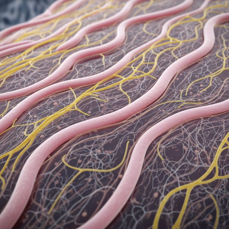
Hyperkeratosis refers to the abnormal thickening of the stratum corneum, the outermost layer of the skin. This condition occurs when the body produces excess keratin, a protective protein found in skin, hair, and nails. The stratum corneum consists of multiple layers of keratinocytes—specialized skin cells that have matured and lost their nucleus and cellular organelles. These flattened, dead cells create a protective barrier against the environment, and when hyperkeratosis develops, this barrier becomes abnormally thick.
The condition manifests in different forms depending on the underlying cause and the nature of keratinocyte maturation. Understanding hyperkeratosis is important because it can indicate various skin conditions ranging from benign mechanical issues to more complex dermatological disorders. While hyperkeratosis itself is not always a disease but rather a histopathological finding, it serves as a clinical indicator of the skin’s response to various stressors.
Types and Classification of Hyperkeratosis
Hyperkeratosis is classified into two main histological categories based on how keratinocytes mature:
- Orthokeratotic Hyperkeratosis: This type involves thickening of the keratin layer while maintaining normal keratinocyte maturation. The keratinocytes properly lose their nuclei as they move toward the skin surface, creating a healthy protective barrier that is simply thicker than normal.
- Parakeratotic Hyperkeratosis: In this form, keratinocytes retain their nuclei, indicating delayed or abnormal maturation. This type is often associated with inflammatory skin conditions and represents a sign that the skin’s normal maturation process has been disrupted.
Additionally, hyperkeratosis can be associated with dyskeratosis, which refers to premature or abnormal keratinization of individual keratinocytes that have not yet reached their normal location in the skin structure. This distinction is important for dermatologists when diagnosing and treating the underlying condition causing hyperkeratosis.
Causes and Etiology of Hyperkeratosis
Hyperkeratosis develops through various exogenous (external) and endogenous (internal) processes. The condition represents the skin’s adaptive response to injury, irritation, or systemic dysfunction.
Mechanical and Chemical Causes
The most common cause of hyperkeratosis in clinical practice is chronic physical damage to the skin. Repeated friction, pressure, or mechanical stress triggers the epidermis to increase its proliferative rate and accelerate keratinocyte maturation. When skin experiences persistent rubbing or pressure, keratinocytes respond by producing more keratin, thereby thickening the stratum corneum. This protective mechanism is evident in conditions such as corns and calluses, which develop on feet and hands from prolonged friction or pressure.
Chemical damage from aggressive soaps and cleansers, particularly those with a basic pH, also contributes to hyperkeratosis development. These harsh products strip the skin of its natural oils and disrupt the skin barrier, prompting a reactive thickening response.
Inflammatory and Systemic Causes
Chronic inflammation represents another significant pathway to hyperkeratosis. Inflammatory skin conditions like psoriasis and dermatitis frequently manifest with hyperkeratotic changes. In these conditions, the immune system’s inflammatory response stimulates keratinocytes to proliferate excessively and produce increased amounts of keratin.
Nutritional deficiencies, particularly vitamin A deficiency, can cause hyperkeratosis through a condition called phrynoderma. Vitamin A plays a crucial role in regulating normal skin cell differentiation and keratinocyte maturation. When deficient, the skin develops characteristic keratin plugs and follicular hyperkeratosis, along with atrophy of sebaceous glands and squamous metaplasia.
Medication-Related Causes
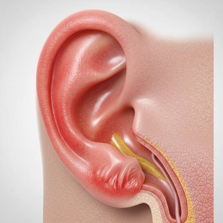
Certain medications can trigger hyperkeratosis as an adverse effect. Chemotherapy drugs and multikinase inhibitors—which target various growth factor receptors including VEGF, PDGFR, EGFR, KIT, RET, and Flt3—frequently cause hyperkeratotic skin reactions. These medications disrupt normal skin homeostasis and can cause characteristic hyperkeratotic hand-foot skin reactions, where hyperkeratosis develops at sites of friction and pressure, particularly on the soles of the feet, causing pain and functional limitation.
Histopathological Features
Different types of hyperkeratosis present distinct histological characteristics that help dermatologists identify the underlying condition:
Mechanical Hyperkeratosis
Mechanical hyperkeratosis is characterized by dense, thickened keratin layers with mild epidermal thickening and variable thickening of the granular layer. Benign, well-defined hyperkeratotic lesions such as corns and calluses show this pattern. Corns are distinguished by a central conical core and are subcategorized into hard corns (heloma durum) and soft corns (heloma molle). The dermis beneath these lesions typically shows collagenization with variable mucin deposition, and inflammatory infiltrates are usually absent.
Psoriasiform Hyperkeratosis
Conditions like psoriasis and psoriasiform dermatitis display characteristic histological features including perivascular aggregates of lymphocytes at the dermal-epidermal junction. These inflammatory cells migrate into the epidermis, causing increased epidermal proliferation and elongation of rete ridges, which creates an undulating or papillomatous appearance. The presence or absence of spongiosis (intercellular edema) helps further differentiate these inflammatory hyperkeratotic conditions.
Symptoms and Clinical Presentation
The symptoms of hyperkeratosis vary significantly depending on its cause and location on the body. Most hyperkeratotic lesions appear as thickened, hardened, or scaly patches of skin. In mechanical hyperkeratosis from corns and calluses, patients typically experience:
- Hardened, raised bumps on feet and hands
- Discomfort or pain, particularly when walking or applying pressure
- Visible thickening and yellowing of the affected skin
- Tenderness when touched
In inflammatory conditions causing hyperkeratosis, symptoms may include itching, redness, flaking, and occasionally pain. When hyperkeratosis is associated with vitamin A deficiency, patients develop keratin plugs that give the skin a characteristic “goose bump” or “toad skin” appearance, particularly on the extensor surfaces of the arms and thighs. Drug-induced hyperkeratosis often develops on pressure-bearing areas and may cause significant functional impairment.
Diagnosis and Evaluation
Diagnosing hyperkeratosis typically begins with a thorough clinical examination and patient history. Dermatologists assess the location, appearance, duration, and associated symptoms. The examination includes close inspection of the affected skin to identify characteristic features and determine the pattern of hyperkeratosis.
In most cases, clinical examination alone is sufficient for diagnosis, particularly when hyperkeratosis is associated with obvious mechanical causes like pressure or friction. However, when the cause is unclear, a skin biopsy may be performed. During a biopsy, a small sample of affected skin is removed and examined under a microscope to identify the specific histopathological pattern, which helps determine the underlying condition.
Additional diagnostic steps may include:
- Detailed assessment of medication history, as certain drugs commonly cause hyperkeratosis
- Evaluation for underlying inflammatory skin conditions
- Nutritional assessment, particularly vitamin A levels if deficiency is suspected
- Dermoscopy, a non-invasive imaging technique that magnifies skin structures
Treatment Options
Treatment of hyperkeratosis depends on identifying and addressing the underlying cause. A multifaceted approach combining causal treatment with symptomatic management typically yields the best results.
Causal Treatment
Eliminating or reducing the causative factor is fundamental to treatment success. For mechanical hyperkeratosis, this involves reducing friction and pressure through proper footwear, custom orthotics, or protective padding. Patients should avoid harsh soaps and switch to gentle, pH-balanced cleansers to prevent chemical irritation. Those with nutritional deficiencies should receive appropriate supplementation, particularly vitamin A when deficiency is confirmed.
Topical Therapies
Keratolytic agents that soften and remove excess keratin are central to symptomatic treatment. Common topical options include:
- Salicylic Acid: This beta-hydroxy acid penetrates the stratum corneum and promotes gentle exfoliation and normalization of keratinization
- Lactic Acid: An alpha-hydroxy acid that hydrates the skin while promoting exfoliation
- Urea: A humectant that increases skin hydration and promotes softer, more pliable skin
- Tretinoin: A retinoid that normalizes keratinocyte differentiation and promotes cellular turnover
Skin Care and Maintenance
Regular moisturization is essential for managing hyperkeratosis. Applying emollients like petroleum jelly, cold cream, or rich moisturizing lotions after bathing helps maintain skin hydration and prevents recurrence. Light exfoliation using pumice stones or gentle scrubs can remove dead skin cells, though aggressive exfoliation should be avoided as it can worsen the condition.
Medical Interventions
For severe or refractory hyperkeratosis, dermatologists may recommend professional treatments such as cryotherapy (freezing), laser therapy, or chemical peels. These interventions remove hyperkeratotic tissue and promote normal skin regeneration.
Prevention Strategies
While some forms of hyperkeratosis cannot be prevented—particularly those with genetic components or certain medication-related causes—several prevention strategies can reduce risk:
- Maintain proper footwear with adequate cushioning and support
- Use gentle, pH-balanced skin care products
- Avoid prolonged friction and pressure on susceptible areas
- Maintain adequate nutrition, particularly vitamin A intake
- Keep skin properly moisturized, especially in dry climates
- Discuss medication side effects with healthcare providers
When to Seek Medical Attention
Consult a dermatologist if:
- Hyperkeratotic lesions are painful or functionally limiting
- The condition worsens despite self-care measures
- New hyperkeratotic lesions develop unexpectedly
- The appearance of lesions changes significantly
- Hyperkeratosis is accompanied by signs of infection
- The condition coincides with starting new medications
Frequently Asked Questions
Q: Is hyperkeratosis permanent?
A: Hyperkeratosis is often manageable rather than permanent. Many cases improve with consistent treatment and elimination of causative factors. However, some forms require ongoing management to prevent recurrence.
Q: Can hyperkeratosis lead to serious complications?
A: Most mechanical hyperkeratosis is benign, though it can cause pain and functional limitations. Hyperkeratosis associated with systemic conditions or certain medications should be evaluated by a healthcare provider to address underlying causes.
Q: Are over-the-counter treatments effective?
A: Many over-the-counter products containing salicylic acid, lactic acid, or urea can help manage mild hyperkeratosis. However, persistent or severe cases benefit from professional evaluation and prescription-strength treatments.
Q: How long does treatment typically take?
A: Response times vary depending on the cause and severity. Mechanical hyperkeratosis may improve within weeks with consistent treatment, while inflammatory or medication-related causes may require longer periods of management.
Q: Can diet affect hyperkeratosis?
A: Yes, adequate nutrition—particularly sufficient vitamin A—is important for normal skin health. Poor nutrition, especially vitamin A deficiency, can contribute to hyperkeratosis development.
References
- Hyperkeratosis — National Center for Biotechnology Information, StatPearls. 2024. https://www.ncbi.nlm.nih.gov/books/NBK562206/
- Keratosis Pilaris (KP) — Massachusetts General Hospital. 2024. https://www.massgeneral.org/condition/keratosis-pilaris
- Hyperkeratosis: Thickening of Keratin in Epidermis — News Medical Life Sciences. 2024. https://www.news-medical.net/health/Hyperkeratosis-Thickening-of-Keratin-in-Epidermis.aspx
Similar Articles


Read full bio of Sneha Tete