Ichthyosis Images: Complete Gallery & Visual Guide
Comprehensive visual guide to ichthyosis types, symptoms, and clinical presentations across various skin conditions.

Ichthyosis refers to a group of genetic skin disorders characterized by dry, thickened, scaly skin resembling fish scales. This article presents a comprehensive collection of clinical images illustrating various types of ichthyosis, aiding in diagnosis and understanding of their presentations. Images depict scaling patterns, severity, and associated features across different forms, from common inherited types to rare congenital variants.
What is ichthyosis?
Ichthyosis encompasses over 20 varieties of cornification disorders where the skin’s outer layer fails to shed properly, leading to persistent dryness, thickening, and scaling. Inherited forms arise from genetic mutations affecting keratinocytes in the stratum corneum, often involving inflammatory pathways like Th17 cytokines. Acquired ichthyosis may signal underlying systemic diseases such as lymphoma or be induced by medications like clofazimine.
Symptoms include fine to plate-like scales, often on extremities, sparing flexures. Severity varies by type, climate, and age; warmer, humid environments typically improve mild cases. Common associations include atopic dermatitis (50% in ichthyosis vulgaris) and increased risks of asthma or allergies.
Ichthyosis vulgaris
The most prevalent inherited ichthyosis, affecting up to 1 in 250 people, ichthyosis vulgaris features fine, white scales on the trunk, arms, and legs, often sparing flexures. It typically manifests in childhood due to filaggrin gene mutations, impairing skin barrier function. Skin appears dry and rough, exacerbated in low humidity.
- Key image descriptions: Close-up of upper arm showing polygonal white scales with underlying erythema; shins with adherent, brownish scales; back with diffuse fine scaling resembling ‘fish skin’.
- Symptoms worsen in winter; mild cases mimic ‘dry skin’. Histology reveals hyperkeratosis and absent granular layer.
Diagnosis is clinical, confirmed by genetic testing for filaggrin mutations via buccal swab. Treatment emphasizes daily emollients (e.g., lanolin-based) and keratolytics for exfoliation.
X-linked ichthyosis
X-linked ichthyosis (XLI), caused by steroid sulfatase deficiency, predominantly affects males. Scales are larger, darker (dirty brown/grey), and ‘adherent’ on neck, trunk, and extremities. Onset occurs in early infancy; corneae may appear cloudy.
- Clinical images: Neck with ‘dirty neck’ scaling; abdomen and thighs covered in polygonal, platelike brown scales; palms showing hyperlinearity.
- Associated with polyhydramnios in pregnancy; mild corneal opacities in 50% of cases.
Diagnostic tests include serum cholesterol sulfate levels and steroid sulfatase assay. Lipoprotein electrophoresis may show abnormalities.
Autosomal recessive congenital ichthyosis (ARCI)
ARCI includes lamellar ichthyosis and congenital ichthyosiform erythroderma (CIE). Neonates present with collodion membrane, evolving into large plate-like scales (lamellar) or erythroderma with fine white scales (CIE). Mutations in transglutaminase-1 or other genes impair barrier formation.
- Image highlights: Lamellar ichthyosis showing thick, dark brown scales on soles, ectropion, and ear scaling; CIE with shiny, red skin and translucent scales; post-collodion phase with fissuring.
- Complications: Ectropion, eclabium, restricted arm movement, infections.
Treatment involves humidified incubators for neonates, topical retinoids, and emollients. Emerging therapies like TMB-001 ointment show promise in reducing scaling.
Lamellar ichthyosis
A severe ARCI subtype, lamellar ichthyosis features prominent palmoplantar keratoderma and large, cracked scales. Skin is taut with potential for secondary infections due to fissures.
- Visual features: Full-body images of ridged, yellow-brown scales; close-ups of heels with hyperkeratosis; scalp involvement with cicatricial alopecia.
Recessive X-linked ichthyosis
Distinct from XLI, this rarer form presents with generalized erythroderma and finer scales, often confused with CIE. Genetic testing differentiates it.
- Images: Widespread fine scaling with underlying redness in infants.
Ichthyosis prematurity syndrome
A rare autosomal recessive disorder where infants are born prematurely (median 32 weeks) encased in thick, clay-like vernix. Post-delivery, respiratory distress is common; survivors have mild scaling lifelong.
- Neonatal images: Thick vernix covering; desquamating skin at 2 weeks; residual mild erythema.
Mothers experience polyhydramnios. Management focuses on respiratory support and emollients.
Acquired ichthyosis
Develops in adulthood, linked to malignancies (e.g., Hodgkin lymphoma), drugs, or autoimmunity. Scales resemble ichthyosis vulgaris but onset is abrupt.
- Presentations: Symmetric scaling on legs in lymphoma patient; drug-induced fine scales.
Complications
Ichthyosis predisposes to infections (bacterial, fungal), dehydration, electrolyte imbalance, and overheating. Severe forms risk sepsis, ectropion, and nail dystrophy. Atopic comorbidities increase in vulgaris and Netherton.
| Complication | Affected Types | Management |
|---|---|---|
| Skin infections | ARCI, HI | Antibiotics, antifungals |
| Dehydration | Neonatal collodion | Humidifiers, fluids |
| Atopic dermatitis | Vulgaris, NS | Topical steroids, dupilumab |
| Skin cancer risk | NS | Regular screening |
Diagnosis
Primarily clinical, supported by family history, biopsy (hyperkeratosis), and genetic testing. Key tests: Skin biopsy, filaggrin mutation analysis, steroid sulfatase activity, serum phytanic acid.
- Histology: Reduced granular layer in vulgaris; electron microscopy shows absent keratohyaline granules.
Treatment
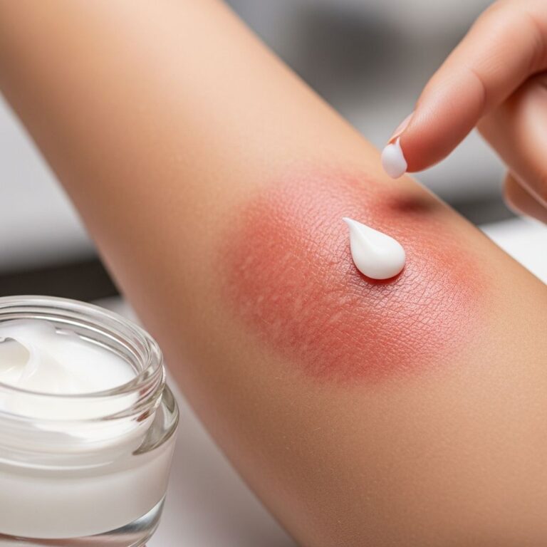
No cure exists; management is symptomatic: daily emollients (3-4x/day), soap substitutes, keratolytics (urea, lactic acid), and exfoliation. Severe cases use oral retinoids. Apply to damp skin post-bath.
- Emollients: High-lipid creams, ointments, bath oils.
- Advanced: Biologics (dupilumab for atopic features), enzyme therapies (TMB-001).
- Avoid soaps, bubble baths.
Podiatry for foot keratoderma; phototherapy (narrowband UVB) for some.
Outlook
Mild forms like vulgaris improve with age and humidity. Severe congenital types persist lifelong but manageable. Early intervention prevents complications; genetic counseling advised.
Frequently Asked Questions (FAQs)
What causes ichthyosis?
Genetic mutations in skin barrier proteins like filaggrin or steroid sulfatase; acquired forms from disease/drugs.
Is ichthyosis contagious?
No, it is genetic or acquired, not infectious.
How is ichthyosis treated in babies?
Humidified incubators, emollients, infection monitoring.
Can ichthyosis be cured?
No, but symptoms improve with consistent moisturizing and keratolytics.
Does ichthyosis improve with age?
Often yes in mild types like vulgaris.
References
- Ichthyosis vulgaris — DermNet NZ. 2023. https://dermnetnz.org/topics/ichthyosis-vulgaris
- Congenital Ichthyosis: A Practical Clinical Guide — PMC/NCBI. 2023-10-01. https://pmc.ncbi.nlm.nih.gov/articles/PMC10503504/
- Living with ichthyosis — Zeroderma. 2023-10. https://www.zeroderma.co.uk/wp-content/uploads/2023/10/Guide-to-ichthyosis-Dec22.pdf
- Ichthyosis — DermNet NZ. 2023. https://dermnetnz.org/topics/ichthyosis
- Ichthyosis – Symptoms, diagnosis and treatment — BMJ Best Practice. 2024. https://bestpractice.bmj.com/topics/en-us/584
- Ichthyosis prematurity syndrome — DermNet NZ. 2023. https://dermnetnz.org/topics/ichthyosis-prematurity-syndrome
Similar Articles


Read full bio of medha deb