Idiopathic Guttate Hypomelanosis Dermoscopy
Detailed dermoscopic insights into idiopathic guttate hypomelanosis, aiding accurate diagnosis of this common hypopigmented skin disorder.

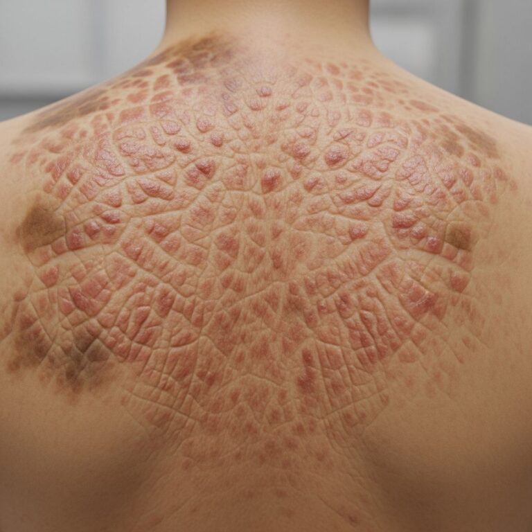
Idiopathic guttate hypomelanosis (IGH) is a common, benign dermatosis characterised by small, round, hypopigmented macules on sun-exposed skin, primarily affecting middle-aged and older adults with fair skin phototypes. Dermoscopy plays a crucial role in confirming the diagnosis by revealing distinctive patterns that differentiate IGH from other hypopigmented lesions.
What is idiopathic guttate hypomelanosis?
Idiopathic guttate hypomelanosis, often abbreviated as IGH, manifests as multiple discrete, white to ivory-coloured spots measuring 2–5 mm in diameter. These lesions are typically asymptomatic and persist lifelong once developed. The term ‘guttate’ refers to their drop-like appearance, while ‘hypomelanosis’ indicates reduced skin pigmentation. IGH predominantly occurs on the extensor surfaces of forearms and shins, reflecting chronic sun exposure as a key aetiological factor.
Clinically, IGH lesions are smooth, well-defined macules without scale, atrophy, or induration. They are more prevalent in females and individuals with Fitzpatrick skin types I–III. Onset usually begins after age 40, coinciding with cumulative UV damage. Although benign, IGH can cause cosmetic distress, prompting patients to seek dermatological advice.
Who gets idiopathic guttate hypomelanosis?
IGH affects approximately 30–50% of fair-skinned individuals over 40 years, with higher prevalence in women (up to 70% in some studies). Risk factors include:
- Chronic sun exposure without adequate protection.
- Age >40 years.
- Fair skin phototype (Fitzpatrick I–III).
- Female gender (possibly due to oestrogen influences on melanocytes).
- Residence in tropical or high-UV regions.
Rarely reported in darker skin types or children, underscoring the role of phototrauma in pathogenesis.
What causes idiopathic guttate hypomelanosis?
The exact aetiology remains idiopathic, but chronic actinic damage is central. Proposed mechanisms include:
- UV-induced melanocyte damage and apoptosis.
- Reduced melanosome transfer from melanocytes to keratinocytes.
- Basement membrane alterations impairing pigment cell function.
- Genetic predisposition interacting with environmental UV exposure.
Histologically, flattened rete ridges, reduced epidermal melanin, and vacuolar degeneration support photoageing as the primary driver.
What are the clinical features of idiopathic guttate hypomelanosis?
IGH presents with:
- Small (2–5 mm), round or oval, hypopigmented macules.
- Symmetrical distribution on extensor forearms, shins, and dorsal hands/feet.
- Smooth surface without scaling, telangiectasia, or textural change.
- No itch, pain, or inflammation.
- Slow progression with new lesions appearing over years.
Lesions may coalesce into larger patches in advanced cases. Differential diagnoses include vitiligo, post-inflammatory hypopigmentation, tinea versicolor, and lichen sclerosus.
How is idiopathic guttate hypomelanosis diagnosed?
Diagnosis is primarily clinical, supported by dermoscopy and history of sun exposure. Dermoscopy is highly specific, revealing hallmark features that obviate biopsy in typical cases.
Dermoscopy of idiopathic guttate hypomelanosis
Dermoscopy of IGH lesions shows a distinctive triad:
- Amorphous white areas: Large, structureless hyporeflective zones due to epidermal atrophy and absent melanin.
- Absence of pigment network: Loss of normal epidermal melanin pattern.
- Peripheral rim of brown dots/granularity: Residual melanin at lesion edges or perifollicular accentuation.
Additional features include:
- Thin, worm-like vessels (rarely).
- Follicular ostia preservation.
- No comedo-like openings or milia-like cysts, unlike other hypopigmented conditions.
These patterns yield >95% diagnostic accuracy. Polarised dermoscopy enhances white structureless areas visibility.
Dermoscopy image gallery
Typical dermoscopic views demonstrate:
- Central white plaque with peripheral pigment specks.
- Variably sized hypopigmented globules.
- Sharp borders contrasting surrounding normally pigmented skin.
(Note: Images would depict 10x magnified views showing the characteristic white amorphous areas, absent network, and marginal brown dots.)
Histopathology of idiopathic guttate hypomelanosis
Biopsy, if performed, reveals:
- Flattened rete ridges with loss of normal architecture.
- Reduced basal layer melanin (confirmed by Fontana-Masson stain).
- Mild vacuolar basal degeneration.
- Normal or slightly thickened stratum corneum.
- No inflammatory infiltrate or scarring.
Electron microscopy shows decreased, smaller melanosomes and damaged melanocytes, corroborating UV aetiology.
Differential diagnosis of idiopathic guttate hypomelanosis
| Condition | Key Differentiators | Dermoscopy |
|---|---|---|
| Vitiligo | Larger lesions, total pigment loss, possible koebnerisation | Complete white with absent follicular pigment |
| Tinea versicolor | Scaly, trunk predominant, positive KOH | Hyphae/spores, brown dots uniformly |
| Lichen sclerosus | Atrophic, wrinkled, genital involvement | Comedo-like openings, white structureless + vessels |
| Post-inflammatory hypopigmentation | History of inflammation, resolves over time | Extracellular melanin, perifollicular sparing |
| Caused by cosmetics/trauma | Confined to application sites | Variable, often with scale |
Dermoscopy reliably distinguishes IGH in most cases.
Treatment of idiopathic guttate hypomelanosis
No curative treatment exists; management focuses on camouflage and prevention:
- Sun protection: Broad-spectrum SPF50+ daily to halt progression.
- Camouflage: Self-tanning lotions, medical tattoos for focal lesions.
- Topical therapies: Tretinoin 0.05% + hydroquinone 4% may repigment mildly (limited evidence).
- Procedural: Cryotherapy, fractional lasers, microneedling reported with variable success and risk of atrophy/hyperpigmentation.
Patient reassurance is paramount, as IGH is harmless.
Prevention of idiopathic guttate hypomelanosis
Primary prevention emphasises UV protection from young adulthood:
- Daily sunscreen application.
- Protective clothing, hats, UV-blocking shades.
- Avoid peak sun hours (10am–4pm).
- Regular antioxidant supplements (e.g., polypodium leucotomos) may offer adjunctive benefit.
Early intervention limits lesion burden.
Idiopathic guttate hypomelanosis FAQs
Is idiopathic guttate hypomelanosis dangerous?
No, IGH is entirely benign with no malignant potential or systemic associations.
Does idiopathic guttate hypomelanosis spread?
It does not spread person-to-person but new lesions may develop with ongoing sun exposure.
Can idiopathic guttate hypomelanosis be cured?
No cure exists, but progression can be halted with sun protection, and camouflage improves appearance.
Is dermoscopy necessary for IGH diagnosis?
Not always, but it confirms diagnosis noninvasively with high specificity, avoiding unnecessary biopsies.
At what age does IGH typically start?
Usually after 40, but can appear earlier in high-risk individuals.
Related topics
- Hypopigmentation
- Photoageing
- Dermoscopy of pigmentary disorders
- Vitiligo dermoscopy
References
- The structure of normal skin — DermNet NZ. 2007. https://dermnetnz.org/topics/the-structure-of-normal-skin
- The dermoscopy report — DermNet NZ. 2008. https://dermnetnz.org/cme/dermoscopy-course/the-dermoscopy-report
- DermNet NZ homepage — DermNet NZ. Accessed 2026. https://dermnetnz.org
- Terminology in dermatology — DermNet NZ. 1997 (updated). https://dermnetnz.org/topics/terminology
- Structure of the dermis and subcutis — DermNet NZ. 2008. https://dermnetnz.org/cme/principles/structure-of-the-dermis-and-subcutis
- DermNet’s Editorial Process — DermNet NZ. Accessed 2026. https://dermnetnz.org/about/editorial-process
Similar Articles


Read full bio of medha deb