Inflammatory Skin Diseases: 5 Key Histologic Patterns
Comprehensive guide to histological patterns and key features of common inflammatory skin conditions in dermatopathology.

Inflammatory skin diseases represent a broad category of dermatological conditions characterized by distinct histological reaction patterns observable under the microscope. Dermatopathology plays a crucial role in their diagnosis, enabling correlation between clinical presentation and microscopic findings. Common patterns include spongiotic, psoriasiform, interface, granulomatous, and bullous reactions, each associated with specific diseases.
Introduction
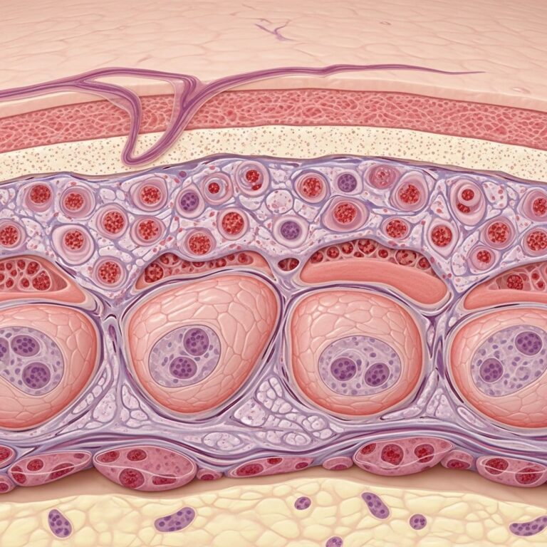
The histopathological evaluation of inflammatory skin diseases involves identifying key reaction patterns that guide diagnosis. These patterns arise from immune-mediated responses targeting the epidermis, dermis, or vascular structures. Accurate biopsy interpretation requires consideration of lesion chronicity, as acute and chronic phases exhibit different features. Direct immunofluorescence (DIF) and special stains often provide confirmatory evidence.
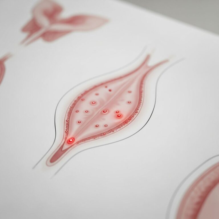
Lesion description is foundational: macules are flat color changes, papules are elevated solid lesions <1 cm, vesicles are fluid-filled <1 cm, and scales indicate retained stratum corneum. Patterns such as perivascular dermatitis (inflammation around vessels) or interface dermatitis (band-like at dermoepidermal junction) help classify diseases.
Eczema
Eczema, or spongiotic dermatitis, is a type IV hypersensitivity reaction mediated by T cells and Langerhans cells responding to allergens. Histologically, it features intercellular edema in the epidermis (spongiosis), leading to vesicle formation in acute phases.
Acute Eczema
Acute lesions show erythematous macules, papules, and vesicles with marked spongiosis, exocytosis of lymphocytes, and occasional Langerhans cell microabscesses. The dermis exhibits superficial perivascular lymphocytic infiltrate. Vesicles contain plasma and inflammatory cells, correlating clinically with weeping bullae.
Subacute Eczema
Subacute phases display diminishing spongiosis, parakeratosis, and acanthosis. Inflammation remains perivascular with some papillary dermal edema.
Chronic Eczema
Chronic eczema evolves to lichenification with psoriasiform hyperplasia, hyperkeratosis, and fibrosis in the papillary dermis. Spongiosis diminishes, and inflammation becomes chronic.
Examples include atopic dermatitis (atopic triad: eczema, asthma, allergies), allergic contact dermatitis, nummular eczema, and id reactions. Diagnosis: “spongiotic dermatitis consistent with eczematous dermatitis.”
Psoriasis
Psoriasis is a T-cell mediated autoimmune disease driven by Th17 cells producing IL-17, attracting neutrophils. It shows psoriasiform dermatitis with regular epidermal hyperplasia.
Key features: parakeratosis, hypogranulosis, suprapapillary thinning, Munro microabscesses (neutrophils in stratum corneum), and dilated papillary vessels. The epidermis is hyperplastic yet focally thinned due to keratinocyte damage. Perivascular lymphocytic infiltrate with neutrophils in severe cases (pustulation).
Overexpressed keratins 6 and 17 reflect hyperproliferation. Clinically, plaques with scale on extensor surfaces.
Lichen Planus
Lichen planus exemplifies interface dermatitis with vacuolar degeneration at the dermoepidermal junction. It features a band-like lymphocytic infiltrate “hugging” the basal layer, basal keratinocyte hydropic change, and Civatte bodies (necrotic keratinocytes).
Epidermis shows compact orthokeratosis, hypergranulosis, and saw-tooth rete ridges. DIF reveals fibrinogen in colloid bodies, occasionally IgM and complement—non-diagnostic as similar in lupus or erythema multiforme.
Variants include hypertrophic and atrophic forms. Clinically, polygonal purple papules (5 Ps: purple, polygonal, pruritic, planar, Wickham striae).
Bullous Pemphigoid
Bullous pemphigoid is the most common subepidermal blistering disease, autoimmune against BP180 and BP230 antigens in the basement membrane.
Histology: subepidermal blister with eosinophils, mast cells, and neutrophils in dermal papillae. Early lesions show eosinophilic microabscesses and perivascular infiltrate.
DIF: linear IgG and C3 at basement membrane zone (BMZ). Salt-split skin shows immunoreactants on the blister roof. Indirect immunofluorescence and ELISA confirm circulating antibodies to 180/230 kD antigens.
Clinically, tense bullae on urticarial plaques in elderly patients.
Vasculitis
Vasculitis involves pathological changes in blood vessels due to immune complex deposition, classified as leukocytoclastic (neutrophilic) or lymphocytic. Features: fibrinoid necrosis, endothelial swelling, karyorrhexis, and red cell extravasation.
Superficial or deep, affects post-capillary venules. Examples: Henoch-Schönlein purpura (IgA), urticarial vasculitis. Erythema elevatum diutinum shows thickened vessels with hemosiderin.
Granuloma Annulare
Granuloma annulare displays palisading granulomas with necrobiosis (collagen degeneration), mucin, and histiocytes in the dermis. Variants: localized, generalized, perforating.
No organisms; interstitial mucin highlighted by Alcian blue. Clinically, annular plaques on extremities.
Acne
Acne vulgaris involves follicular rupture leading to mixed inflammation: neutrophils acutely, lymphocytes chronically, and foreign body giant cells. Features: dilated follicles with keratin, bacteria (Propionibacterium acnes), perifollicular abscesses, and granulomatous reactions in nodulocystic forms.
Comedones show basket-weave orthokeratosis.
Other Patterns
- Interface Dermatitis: Vacuolar (lupus, dermatomyositis) vs. lichenoid (lichen planus). Basal vacuolization, necrotic keratinocytes.
- Granulomatous: Discrete histiocytic collections; caseating (TB, Fite+), non-caseating (sarcoid). Rosacea: perifollicular granulomas.
- Dermatitis Herpetiformis: Subepidermal split, papillary microabscesses, granular IgA on DIF; celiac association.
- Lupus Erythematosus: Interface with atrophy, basement membrane thickening, dermal mucin. DIF: granular IgG/M/C3 at BMZ.
Frequently Asked Questions (FAQs)
Q: What is the hallmark of spongiotic dermatitis?
A: Intercellular edema (spongiosis) in the epidermis with lymphocytic exocytosis, seen in eczema.
Q: How does psoriasis differ histologically from eczema?
A: Psoriasis shows psoriasiform hyperplasia, parakeratosis, Munro abscesses, and suprapapillary thinning; eczema has spongiosis.
Q: What DIF finding confirms bullous pemphigoid?
A: Linear IgG and C3 along the BMZ, roof-binding on salt-split skin.
Q: Describe lichen planus histology.
A: Band-like infiltrate, basal vacuolization, Civatte bodies, hypergranulosis.
Q: What are key features of leukocytoclastic vasculitis?
A: Fibrinoid necrosis, endothelial swelling, neutrophilic karyorrhexis, RBC extravasation.
| Pattern | Key Features | Examples |
|---|---|---|
| Spongiotic | Spongiosis, vesicles, perivascular lymphs | Eczema, contact dermatitis |
| Psoriasiform | Hyperplasia, parakeratosis, Munro abscesses | Psoriasis |
| Interface | Vacuolar change, band-like infiltrate | Lichen planus, lupus |
| Bullous | Subepidermal split, eosinophils | Bullous pemphigoid |
| Granulomatous | Histiocytes, necrobiosis, mucin | Granuloma annulare |
References
- Introduction to Inflammatory Dermpath — Schaberg, UC Davis. 2019-07-01. https://schaberg.faculty.ucdavis.edu/wp-content/uploads/sites/604/2019/07/Inflammatory_Derm_handout.pdf
- INFLAMMATORY DISEASES OF THE SKIN — Duke Pathology. 2021-06-21. https://pathology.oit.duke.edu/siteParts/avaps/06.21.1_Inflammatory_Diseases_of_the_Skin_Final.pdf
- Inflammatory skin diseases – Dermatopathology — DermNet NZ. Accessed 2026. https://dermnetnz.org/cme/dermatopathology/inflammatory-skin-diseases
- Dermatopathology 101: Part 1 – Inflammatory skin diseases — PubMed/NCBI. 2017-02-01. https://pubmed.ncbi.nlm.nih.gov/28140531/
- Introduction to the Dermatopathology of Inflammatory Skin Diseases — Weill Cornell Medicine YouTube. 2018. https://www.youtube.com/watch?v=0FDa5S9g-18
Similar Articles


Read full bio of medha deb