Iris: Anatomy, Function & Eye Health Guide
Complete guide to iris anatomy, function, and common conditions affecting your eye's colored guardian.

The iris is one of the most visible and fascinating parts of your eye. This colored, circular structure plays a crucial role in your vision by controlling how much light enters your eye. Beyond its essential function in regulating light, the iris also determines your eye color and contributes to your unique appearance. Understanding the anatomy and function of the iris can help you better appreciate how your eyes work and recognize when something might be wrong.
What Is the Iris?
The iris is the colored muscular part of the eye surrounding the pupil, the small black opening in the center of your eye. Located in front of the lens and behind the cornea, the iris is bathed in a clear fluid known as aqueous humor, which nourishes and protects the eye. The iris functions as your eye’s light controller, automatically adjusting to changing light conditions to protect your retina and ensure optimal vision.
Many people confuse the iris with other parts of the eye. The iris and retina are distinctly different structures. While the iris regulates light entering the eye, the retina is a layer of tissue at the back of the eye that translates light into images. These two structures work together harmoniously to enable clear vision.
Iris Anatomy and Structure
Location in the Eye
The iris is located in the centre of the eye, surrounding the pupil, positioned under the cornea and on top of the lens. This strategic location allows the iris to act as a gatekeeper, controlling exactly how much light reaches the sensitive tissues behind it. The iris is essentially one layer from the outside of the eyeball, making it both visible and functionally critical to your vision.
Structural Composition
The iris is composed of several distinct layers, each serving specific purposes in eye function:
- Stromal Layer: The visible colored portion of the iris that contains connective tissue and blood vessels. The amount of pigment in this layer determines your eye color.
- Muscular Layer: Comprises two groups of muscles—the dilator pupillae and sphincter pupillae—that control pupil size in response to light. These smooth muscles work in opposing directions to adjust the pupil opening.
- Posterior Pigmented Epithelium: A darkly pigmented layer that prevents light from passing through the iris, ensuring light enters only through the pupil.
Two Regions of the Iris
The iris is divided into two regions: the pupillary zone and the ciliary zone. The pupillary zone is the inner region located at the margin of the pupil, known as the pupillary ruff. The ciliary zone is the larger outer region adjacent to the ciliary body. These two zones join at the collarette, which consists of nerves and blood vessels that support iris function.
How the Iris Controls Light
The Pupillary Light Reflex
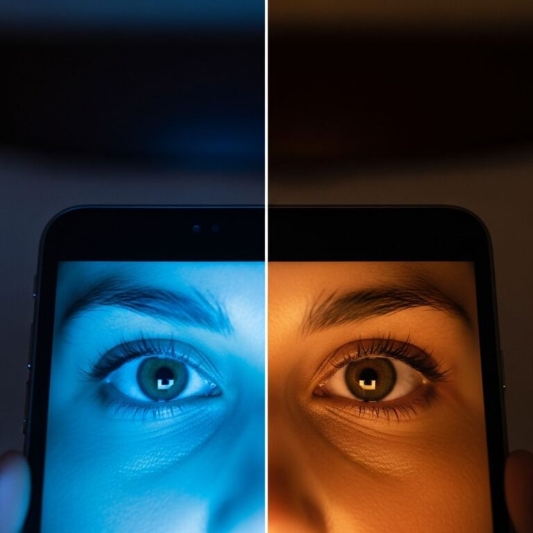
The primary function of the iris is to control the diameter and size of the pupil, thereby regulating the amount of light that reaches the retina. This automatic process is known as the pupillary light reflex. Your iris responds instantaneously to changes in lighting conditions, protecting your eye from excessive light and maximizing vision in darkness.
How Muscles Control Pupil Size
Two distinct muscles in your iris control how your pupil opens and closes:
- Sphincter Muscle: This circular muscle contracts when exposed to bright light, causing the pupil to constrict and reduce the amount of light entering the eye. When the sphincter contracts, it narrows the pupil opening significantly.
- Dilator Muscle: This muscle expands the pupil opening when it contracts, allowing more light to enter in low light conditions. The dilator muscle looks somewhat like a sun with rays extending outward from the center.
When you move from a bright room into darkness, your dilator pupillae muscle contracts to dilate your pupil and allow more light in. Conversely, when exposed to bright light, your sphincter pupillae muscle contracts to constrict your pupil and reduce light entry. This automatic adjustment happens without conscious thought, protecting your retina from damage while maintaining optimal vision.
Eye Color: How the Iris Determines Your Look
Your eye color is determined by the amount and type of pigment contained in the iris. This pigmentation in the stromal layer of the iris creates the wide spectrum of eye colors we see in the human population.
The Pigment Scale
Eye color follows a relatively simple rule based on melanin pigmentation:
- Blue Eyes: When there is very little pigment in the iris, the eye appears blue.
- Green to Hazel Eyes: Moderate amounts of pigment produce green and hazel eye colors.
- Brown to Black Eyes: With increased pigment, the shade becomes deep brown to black.
Eye color is primarily determined by genetics, with multiple genes influencing melanin production and distribution in the iris. While brown eyes are the most common eye color worldwide due to higher melanin content, blue eyes are more common in Northern European populations where lower melanin levels are more prevalent.
The Iris and Vision
How the Iris Helps You See
The iris helps the human eye see by controlling how much light the pupil lets in. The complete vision process involves multiple eye structures working together. The cornea allows light to enter the eye, the iris adapts the pupil according to light intensity, the lens directs and concentrates light onto the retina, the retina receives focused light, and the optic nerve carries visual information to the brain.
Without the iris’s ability to regulate light, your vision would be severely compromised. In bright conditions, excess light could damage your retina. In dim conditions, insufficient light would reach your retina, making vision impossible. The iris’s automatic adjustment maintains the perfect balance for optimal vision across all lighting conditions.
Connection to Other Eye Structures
The iris, ciliary body, and choroid are connected and make up an area of your eye called the uvea. The choroid is a network of blood vessels located at the back of your eye between your sclera and retina. The ciliary body connects on each side of your iris to control the movement of your lens. These structures work together seamlessly, with the iris and ciliary body flowing into each other like one curved street that changes names and function as it progresses around the eye.
Common Iris-Related Conditions
Several conditions can affect the iris and impact your vision and eye health:
- Iritis (Anterior Uveitis): Inflammation of the iris that commonly has no determinable cause. As a result of inflammation, the iris may stick to the lens or cornea, blocking the normal flow of fluid in the eye.
- Glaucoma: Can result as a complication of iris inflammation or from other iris-related issues.
- Aniridia: A rare condition involving partial or complete absence of the iris.
- Coloboma: A congenital condition where part of the iris is missing, creating an unusual pupil shape.
- Pigment Dispersion Syndrome: A condition where iris pigment is released into the eye.
- Horner’s Syndrome: A neurological condition affecting iris function and appearance.
Treatment for Iris Conditions
Complications of iritis include secondary glaucoma and blindness, making proper treatment essential. Treatment for iris inflammation usually involves topical steroid eye drops to reduce inflammation and prevent complications. Your eye care provider may also use eye drops that paralyze the ciliary body to test you for refractive errors such as nearsightedness or farsightedness.
Maintaining Iris and Eye Health
Protective Measures
While you cannot directly control iris function, you can protect your eyes and iris health through several measures:
- Wear UV-protective sunglasses to protect your iris from harmful ultraviolet radiation
- Avoid prolonged exposure to extremely bright light or laser light
- Protect your eyes from trauma and injury
- Maintain proper eye hygiene to prevent infections
- Get regular eye exams to monitor iris and overall eye health
When to See an Eye Care Provider
Contact your eye care provider if you experience:
- Eye pain or severe discomfort
- Redness or swelling around the iris area
- Changes in pupil size or responsiveness to light
- Blurred vision or difficulty with light sensitivity
- Any unusual changes in your iris appearance
Frequently Asked Questions About the Iris
Q: Can the iris change color?
A: In rare cases, iris color can change due to certain medications, eye injuries, or medical conditions like Horner’s syndrome. However, eye color is primarily determined by genetics and remains relatively stable throughout your life.
Q: Why do my pupils dilate in darkness?
A: Your pupils dilate (enlarge) in darkness because your dilator pupillae muscle contracts, allowing more light to reach your retina. This automatic response helps you see better in dim lighting conditions.
Q: Is the iris visible without looking in a mirror?
A: Yes, the iris is the visible colored part of your eye that others can see when they look at your face. It surrounds the pupil and is clearly visible from the front.
Q: What does it mean if my iris appears to have different colors?
A: Some people have heterochromia, a condition where each iris contains different amounts of pigment, resulting in different colored eyes. This is usually harmless and can be genetic or result from injury or certain medical conditions.
Q: Can iris problems affect my vision?
A: Yes, serious iris conditions like iritis or coloboma can affect your vision. Inflammation can block normal fluid flow in the eye, and structural abnormalities can affect light regulation. Proper diagnosis and treatment are essential.
Q: How fast do pupils respond to light changes?
A: Your pupils typically respond to light changes within milliseconds. This rapid response is an automatic reflex controlled by your iris muscles, protecting your eye from light damage while maximizing vision.
References
- Marvels of the Iris: Understanding the Eye’s Colorful Guardian — University of Ophthalmology and Optometry. 2024. https://www.uoosd.com/marvels-of-the-iris
- Ciliary Body of the Eye: Anatomy and Function — Cleveland Clinic. 2024. https://my.clevelandclinic.org/health/body/24839-ciliary-body
- Iris: Anatomy, Function, and Iris-related Conditions — Oscar Wylee Eye Care. 2024. https://www.oscarwylee.com.au/glasses/eye/anatomy/iris
- Iris: Eye, Structure, Anatomy, & Function — Britannica. October 13, 2025. https://www.britannica.com/science/iris-eye
- Pupil of the Eye: Definition, Anatomy & Function — Cleveland Clinic. 2024. https://my.clevelandclinic.org/health/body/24317-pupil-of-the-eye
- Eye Anatomy Explained by Experts — Cannon EyeCare. 2024. https://seattleeyecaredoctor.com/eye-anatomy-101/
- Anatomy of the Eye — Cleveland Eye Clinic. February 23, 2024. https://clevelandeyeclinic.com/2024/02/23/anatomy-of-the-eye/
Similar Articles


Read full bio of medha deb