Jessner Lymphocytic Infiltrate: Comprehensive Guide
Understanding Jessner lymphocytic infiltrate: a benign T-cell skin disorder with red papules on sun-exposed areas.

Jessner lymphocytic infiltrate (JLIS), also known as Jessner’s lymphocytic infiltration of the skin, is a rare, benign, chronic T-cell lymphoproliferative disorder characterized by recurrent red papules and plaques primarily on sun-exposed areas.
What is Jessner Lymphocytic Infiltrate?
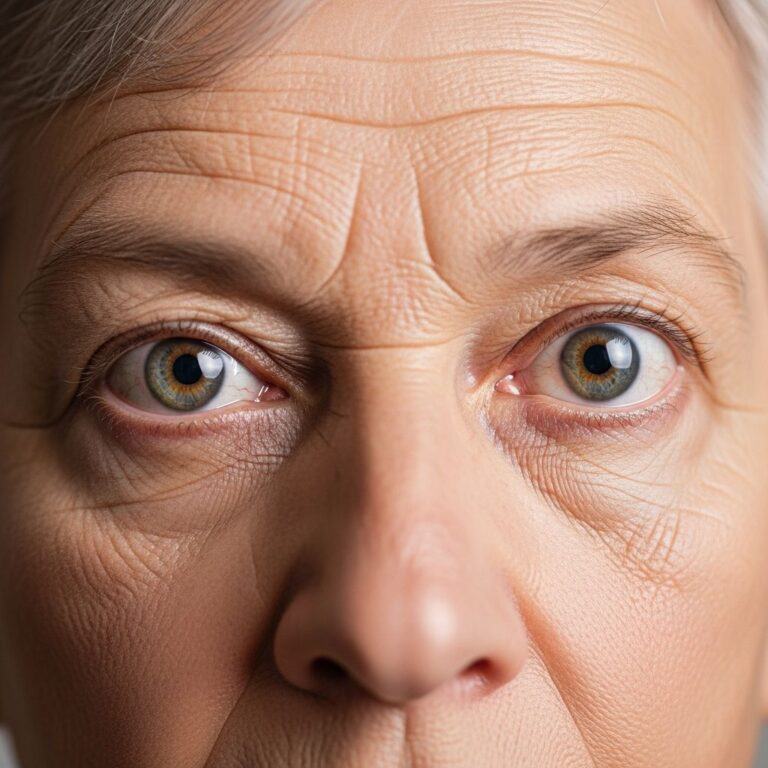
Jessner lymphocytic infiltrate is an idiopathic skin condition first described in 1953 by Max Jessner and colleagues. It manifests as asymptomatic or mildly pruritic lesions that appear as non-scaly, reddish papules, nodules, or plaques, typically on the face, neck, upper back, and shoulders—regions most exposed to sunlight. Unlike malignant lymphomas, JLIS is self-limited and benign, though it can persist for years with periods of remission and recurrence.
The condition involves a dense infiltrate of lymphocytes in the dermis without epidermal involvement, distinguishing it from other dermatoses. Recent immunohistochemical studies confirm a predominance of T-lymphocytes, refuting earlier notions of it being a pseudo-B-cell lymphoma akin to lymphocytoma cutis.
Who Gets Jessner Lymphocytic Infiltrate?
JLIS most commonly affects adults aged 30–60 years, with no strong gender predominance, though some studies report slight female preponderance (e.g., 54 females in a cohort of 100 patients). It is rare in children and the elderly, but cases like a 73-year-old woman have been documented.
There is no established ethnic predilection, but photosensitivity suggests a role for ultraviolet light exposure, potentially explaining higher incidence in fair-skinned individuals living in sunny climates.
Clinical Features
Lesions typically present as:
- Erythematous papules: Small, red, dome-shaped bumps, 2–5 mm in diameter, non-scaly and firm to touch.
- Plaques: Annular or discoid patches up to 1–2 cm, often with central clearing, expanding peripherally.
- Nodules: Larger, deeper lesions in chronic cases.
Skin surrounding lesions may show erythema or mild scaling. Symptoms are usually asymptomatic, but pruritus, burning, or tenderness can occur, especially with sun exposure. Photosensitivity is common, with lesions worsening after UV exposure.
Distribution
Predominantly photo-exposed sites: face (malar cheeks), neck, ears, upper chest, back, and arms. Lesions spare covered areas like the trunk below the waist.
Histology and Pathophysiology
Histologically, JLIS shows a dense, band-like perivascular and periadnexal lymphocytic infiltrate in the upper and mid-dermis, sparing the epidermis and deep dermis. Key features include:
- Predominance of CD4+ T-cells with few B-cells or plasma cells.
- No atypia, epidermotropism, or mucin deposition (unlike lupus).
- Mild dermal edema in early lesions; fibrosis in chronic ones.
Immunohistochemistry on frozen sections confirms T-cell dominance, differentiating it from B-cell pseudolymphomas. Pathogenesis is unclear but may involve aberrant T-cell response to UV light or autoantigens, without systemic involvement.
Differential Diagnosis
JLIS mimics several conditions; biopsy is essential for distinction. Common differentials include:
| Condition | Key Distinguishing Features |
|---|---|
| Polymorphous Light Eruption (PMLE) | More pruritic, polymorphic lesions; resolves faster; positive phototesting. |
| Discoid Lupus Erythematosus (DLE) | Scaly, atrophic scars, interface dermatitis, mucin; positive ANA possible. |
| Lupus Erythematosus Tumidus (LET) | Urticarial plaques with mucin; DEJ changes; may overlap spectrum with JLIS. |
| Lymphocytoma Cutis | B-cell predominant; nodular; responds to antimalarials. |
| Cutaneous T-cell Lymphoma (CTCL) | Atypical lymphocytes, epidermotropism, progression. |
| Drug Eruption | History of new medication; resolves on discontinuation. |
A study of 100 patients confirmed JLIS as a distinct entity without progression to DLE, PMLE, or lymphoma, though coexistence with PMLE in 10% was noted.
Diagnosis
Diagnosis relies on:
- Clinical presentation: Photo-distributed red papules/plaques.
- Skin biopsy: Confirmatory with dense dermal T-lymphocytic infiltrate.
- Laboratory tests: Normal blood work, negative ANA, rule out infection/malignancy.
- Phototesting: Optional to assess photosensitivity.
Immunohistochemistry (CD3+, CD4+, CD20-) and absence of clonality distinguish benign JLIS from lymphoma.
Treatment
No curative treatment exists; management focuses on symptom control and sun protection. First-line approaches:
- Sun protection: Broad-spectrum SPF 50+ sunscreen reapplied every 2–3 hours; hats, UPF clothing.
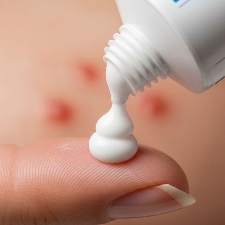
- Topical corticosteroids: Potent agents (e.g., clobetasol) intermittently for inflammation.
- Intralesional steroids: Triamcinolone 10 mg/mL (0.5 mL per lesion), effective with complete resolution in cases.
Second-line options for refractory cases:
- Antimalarials (hydroxychloroquine 200–400 mg/day).
- Topical calcineurin inhibitors (tacrolimus).
- Systemic steroids or immunosuppressants rarely.
In a case report, intralesional triamcinolone twice over 15 days plus sunscreen led to full resolution without recurrence at 5-month follow-up. Many patients experience spontaneous remission after years.
Prognosis and Complications
JLIS is benign with no malignant potential or systemic involvement. Lesions wax and wane, often remitting after 5–10 years. Post-inflammatory hyperpigmentation may occur but fades with sun protection. Rare scarring if untreated.
Prevention
Daily photoprotection is key: mineral-based SPF 50+ sunscreen, avoiding peak sun hours (10 AM–4 PM), and protective clothing prevent flares.
Frequently Asked Questions (FAQs)
Q: Is Jessner lymphocytic infiltrate cancerous?
A: No, it is a benign T-cell lymphoproliferative disorder without risk of progression to lymphoma.
Q: Does sun exposure worsen Jessner lymphocytic infiltrate?
A: Yes, lesions often flare with UV exposure; strict sun protection is essential.
Q: How long do lesions last?
A: Individual lesions persist months; the condition may last years but often remits spontaneously.
Q: Can Jessner lymphocytic infiltrate be cured?
A: No cure, but treatments like intralesional steroids and sun protection achieve long-term control.
Q: Is biopsy always needed?
A: Yes, to confirm diagnosis and exclude mimics like lupus or lymphoma.
References
- Lymphocytic infiltration of the skin (Jessner) — British Journal of Dermatology. 1984-05-01. https://academic.oup.com/bjd/article/110/5/523/6689292
- Jessner’s Lymphocytic Infiltration of the Skin in a 73-Year-Old Woman — American Journal of Case Reports. 2021-01-01. https://amjcaserep.com/abstract/full/idArt/938969
- Jessner’s Lymphocytic Infiltration of the Skin: A Clinical Study of 100 Patients — JAMA Dermatology. 1992-06-01. https://jamanetwork.com/journals/jamadermatology/fullarticle/551077
- Lymphocytic Infiltrate of Jessner — National Organization for Rare Disorders (NORD). 2023-01-01. https://rarediseases.org/rare-diseases/lymphocytic-infiltrate-of-jessner/
- Jessner’s Lymphocytic Infiltrate — Primary Care Dermatology Society (PCDS). 2021-08-08. https://www.pcds.org.uk/clinical-guidance/jessners-lymphocytic-infiltrate
- Lupus Erythematosus Tumidus vs Jessner’s Lymphocytic Infiltrate — Practical Dermatology. 2023-01-01. https://practicaldermatology.com/topics/general-topics/lupus-erythematosus-tumidus-vs-jessners-lymphocytic-infiltrate-of-the-skin/21686/
- Jessner Lymphocytic Infiltration of the Skin — StatPearls, NCBI Bookshelf. 2023-06-26. https://www.ncbi.nlm.nih.gov/books/NBK562281/
Similar Articles


Read full bio of Sneha Tete